This is especially significant given that a 2009 study by Lai et al.2 found that more than 90% of surgeons performing AUS implants conducted five or fewer cases per year (median ≈ 1 to 2), and a 2016 review by Cordon et al.3 reported that only about 4% of urologists were considered “high-volume” AUS implanters (defined as > 20 cases/year).
Beyond the implantation itself, evaluating a patient who presents with recurrent incontinence after a seemingly successful AUS procedure can be one of the more complex and uncertain aspects of AUS management. The potential causes of recurrent incontinence are numerous—mechanical failure, urethral atrophy, cuff erosion, or infection—and standard diagnostic tools such as history, physical examination, and office cystoscopy are often inconclusive.
A recent publication in Investigative and Clinical Urology describes a practical and durable solution to this diagnostic challenge: filling the AUS system with iso-osmotic contrast at the time of implantation. This simple modification, pioneered at Mayo Clinic in the 1970s and continued through today, allows straightforward radiographic evaluation of any patient presenting with recurrent stress incontinence.
When the system is filled with contrast, a single plain pelvic X-ray can distinguish between an intact system, an early leak, or complete mechanical failure—without the need for CT, ultrasound, or surgical exploration. A smooth, round reservoir indicates integrity; a partially collapsed or irregular contour indicates early leakage; and the absence of contrast confirms mechanical failure. This rapid, inexpensive, and definitive test transforms what was once a diagnostic gray zone into a clear, reproducible algorithm.
The Mayo institutional experience spans more than 3,000 AUS surgeries—the largest single-center series in the world. Within this database, over 2,000 primary implants have been filled with iso-osmotic contrast rather than saline. Across this extensive cohort, the use of contrast has proven entirely safe, with no increase in mechanical failure rates, infection, or device revisions. Mechanical failure occurred in 13.5% of patients, with a median time to failure of 4.9 years and nearly 90% device survival at five years—comparable to or better than rates reported in large multi-institutional reviews.
Importantly, every case of mechanical failure identified radiographically was confirmed preoperatively, underscoring the accuracy of this diagnostic method. The simplicity of the technique also facilitates standardized evaluation by any urologist, including those with less extensive AUS experience. In an operation with such a steep learning curve, this kind of diagnostic aid can be invaluable.
Although Boston Scientific recommends filling the AUS with normal saline, the manufacturer lists several approved contrast alternatives. The preparation used in this series—mixing 48 mL of Omnipaque 350 with 60 mL of sterile water—takes less than one minute of OR time and adds no meaningful operative complexity or cost.
Ultimately, the iso-osmotic contrast technique exemplifies how to overcome diagnostic and technical barriers. For high-volume prosthetic surgeons, it eliminates uncertainty and accelerates revision decision-making. For newer surgeons still in the early stages of mastering the intricacies of AUS management, it provides confidence and diagnostic precision that would otherwise take years to acquire.
Long-term institutional experience demonstrates that iso-osmotic contrast filling is a safe, cost-effective, and simple diagnostic tool that can be considered by surgeons seeking to optimize the care of their AUS patients.
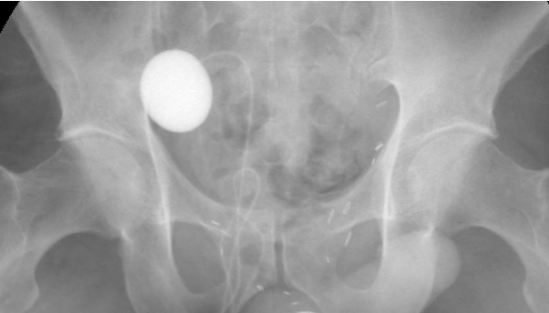
Figure 1: Initial plain film imaging following successful AUS implantation with contrast in the reservoir

Figure 2: Seven years post-op, the patient returns with a new onset of incontinence. Plain film demonstrates a nearly empty reservoir consistent with a mechanical failure of the device
References:
- Sandhu JS, Maschino AC, Vickers AJ. The surgical learning curve for artificial urinary sphincter procedures compared to typical surgeon experience. European Urology. 2011;60(6):1285–1290.
- Lai HH HE, Teh BS, Butler EB, Boone TB. 13 years of experience with artificial urinary sphincter implantation at Baylor College of Medicine. J Urol. 2007;177(3):1021-5.
- Cordon BH, Singla N, Singla AK. Artificial urinary sphincters for male stress urinary incontinence: current perspectives. Medical Devices: Evidence and Research. 2016;9:175–183
Written by: Daniel S. Elliott, MD, and Maraika O. Robinson, MD, Mayo Clinic, Rochester, Minnesota, USA
Read the Abstract