(UroToday.com) The 2025 SUO annual meeting featured a prostate cancer session and a presentation by Dr. Rana McKay discussing validation of multimodal and image-only artificial intelligence digital pathology-based biomarkers using multi-institutional real-world data. Artificial intelligence digital pathology-based biomarkers are gaining adoption in clinical practice for their ability to practically and efficiently personalize patient care. At SUO 2025, Dr. McKay and colleagues further demonstrated the value of digital pathology through validation of:
- A multimodal artificial intelligence biomarker utilizing digital pathology images and key clinical variables (age, PSA, T-stage)
- A newly developed “image-only” artificial intelligence biomarker utilizing only digital pathology images
The investigators assessed both models for prediction of the primary outcome of 10-year risk of distant metastasis and secondary outcome of prostate cancer specific mortality in a contemporary cohort of patients with localized prostate cancer treated at three US institutions:
- University of California, San Diego
- University of California, San Francisco
- University of Kansas Medical Center
The multimodal artificial intelligence (version 1.2) and image-only (version 1.3) models were previously trained and locked prior to this multicenter validation study:1

Retrospective clinical data and digital pathology from first available routine prostate biopsies were compiled from each of the participating sites between February 2024 to September 2024. Eligible patients were diagnosed between 2005-2020 and had evaluable digital histopathology and complete clinical data. The association between each biomarker and distant metastasis or prostate cancer specific mortality was evaluated continuously (per standard deviation) and categorically [raw continuous scores, ranging from 0-1, were categorized into 3 risk groups (low, intermediate, high) at the time of development and locked prior to validation] using univariable and multivariable Fine-Gray proportional hazard regression models and cumulative incidence analyses.
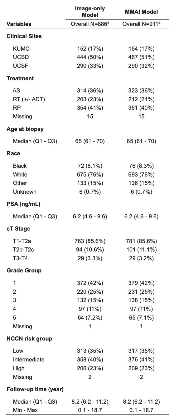
Due to different image pre-processing between the multimodal artificial intelligence and image-only models, multimodal artificial intelligence scores were generated for 991 patients and image-only scores for 886 patients. The multimodal artificial intelligence cohort was composed of 35% NCCN low risk, 41% intermediate risk, and 23% high risk patients with median age of 65 years (IQR 61-70), PSA 6.2 ng/mL (IQR 4.6-9.6), and median follow-up of 8.2 years (IQR 6.2-11.2). Primary treatment included active surveillance (36%), radiation therapy +/- ADT (24%), and prostatectomy (40%). Most patients were white (76%) or black (8.3%), and Grade Group distribution was: 42% Grade Group 1, 25% Grade Group 2, 15% Grade Group 3, 11% Grade Group 4, and 7.1% Grade Group 5. Baseline characteristics were similar to the image-only cohort:
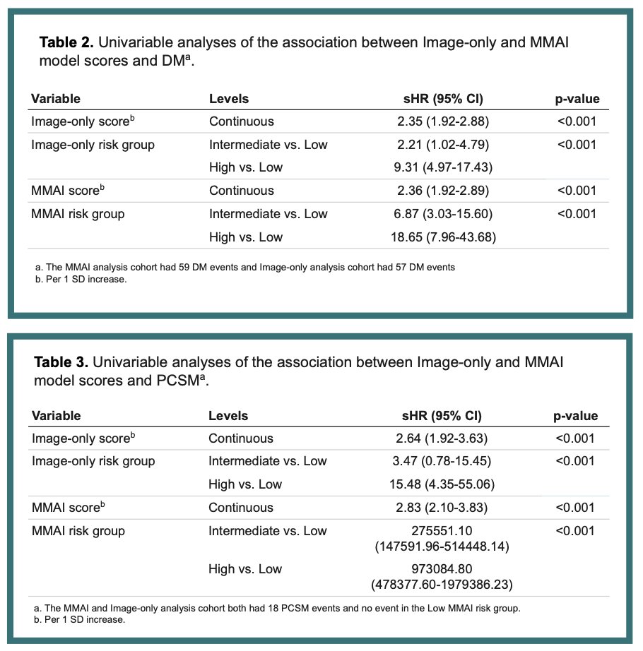
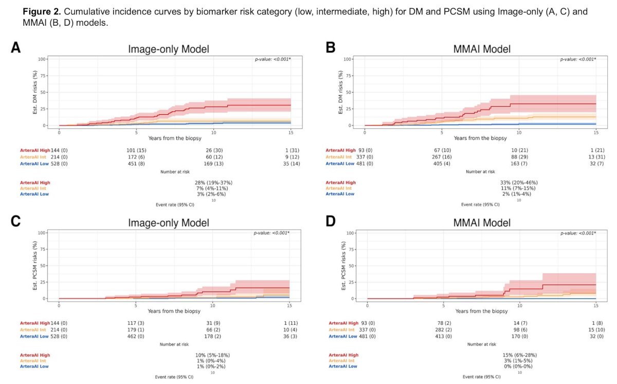
Both multimodal artificial intelligence and image-only continuous and categorical scores were significantly associated with 10-year risk of distant metastasis and prostate cancer specific mortality:
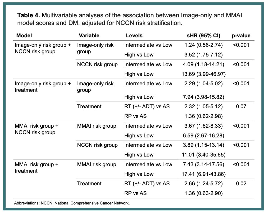
Both models also remained significant when controlling for NCCN risk group or treatment:
The models had similar categorical risk distribution and there were no notable differences in performance by race (results not shown):
Dr. McKay concluded her presentation discussing validation of multimodal and image-only artificial intelligence digital pathology-based biomarkers using multi-institutional real-world data with the following take home points:
- This study confirms the generalizability and prognostic utility of the multimodal artificial intelligence model in contemporary United States clinical settings
- While the multimodal artificial intelligence model has been validated in numerous other studies, this is the first validation of an image-only model (not commercially available, as of yet), which offers similar prognostic ability
- These findings substantiate the presence of valuable prognostic information within histopathology and suggest promise for scalable, image-based risk stratification that does not require clinical inputs
Presented by: Rana McKay, MD, UC San Diego, San Diego, CA
Written by: Zachary Klaassen, MD, MSc – Urologic Oncologist, Associate Professor of Urology, Georgia Cancer Center, Wellstar MCG Health, @zklaassen_md on Twitter during the 2025 Society of Urologic Oncology (SUO) annual meeting held in Phoenix, AZ, between the 2nd and 5th of December 2025.
References:
- Gerrard P, Zhang J, Yamashita R, et al. Analytical validation of a clinical grade prognostic and classification artificial intelligence laboratory test for men with prostate cancer. AI in Precision Oncology. 2024;1(2).