(UroToday.com) The 2026 SNMMI annual meeting featured a genitourinary diagnosis and imaging session and a presentation by Liang Zhao, MD, PhD, discussing a head-to-head comparison of 68Ga-OncoACP3 and 68Ga-PSMA-11 PET for imaging prostate cancer. Radiopharmaceuticals targeting PSMA are the cornerstone of prostate cancer imaging and theranostics. However, a substantial proportion of patients and tumor lesions exhibit low or absent PSMA expression, limiting the sensitivity and clinical applicability of PSMA-based agents. Prostatic acid phosphatase (ACP3) is expressed in a higher proportion of prostate cancers while being virtually absent from most normal tissues, making it a potentially superior diagnostic and therapeutic target. A novel ACP3-targeting ligand, OncoACP3, developed by Georgiev et al.1, has demonstrated high tumor uptake and specificity in preclinical prostate cancer models, supporting its clinical translation. This study aimed to systematically evaluate 68Ga-OncoACP3 for in-human ACP3 PET imaging and to perform a head-to-head comparison with 68Ga-PSMA-11.
This prospective clinical study (NCT07242014) enrolled patients with newly diagnosed prostate cancer, biochemical recurrence, or treatment-refractory disease who underwent paired 68Ga-OncoACP3 and 68Ga-PSMA-11 PET/CT scans between September and December 2025. Wilcoxon rank-sum tests were used to compare normal-organ biodistribution and tumor uptake between the two tracers, and lesion detection performance was assessed. In a subset of patients, dual-time-point 68Ga-OncoACP3 imaging at 1 and 2 hours post-injection was performed to evaluate short-term uptake kinetics.
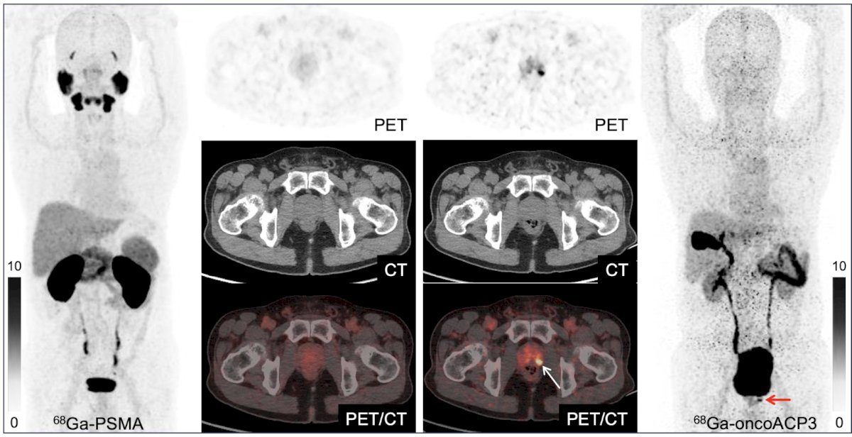
A total of 65 patients were enrolled, including 51 with newly diagnosed disease, 6 with biochemical recurrence, and 8 with treatment-progressive prostate cancer. 68Ga-OncoACP3 demonstrated significantly lower physiologic uptake than 68Ga-PSMA-11 in the lacrimal glands, parotid glands, submandibular glands, sublingual glands, liver, spleen, blood pool, and kidneys (all p < 0.001), with the most pronounced difference observed in the kidneys (median SUVmean 4.1 [IQR 3.3–5.0] versus 27.7 [IQR 21.9–33.6], p < 0.001). Due to hepatobiliary excretion, 68Ga-OncoACP3 showed significantly higher uptake in the biliary system (median SUVmean 16.2 [IQR 12.3–27.0] versus 2.9 [IQR 1.9–5.6], p < 0.001). Tumor uptake of 68Ga-OncoACP3 was significantly higher than that of 68Ga-PSMA-11 in primary prostate tumors (SUVmax 22.0 [IQR 13.2–37.0] versus 14.8 [IQR 7.4–24.5], p < 0.001):
In patients with treatment-refractory disease, metastatic lymph nodes also showed significantly higher uptake with 68Ga-OncoACP (SUVmax 14.7 [IQR 7.5–26.6] versus 7.2 [IQR 3.0–18.1], p < 0.001):
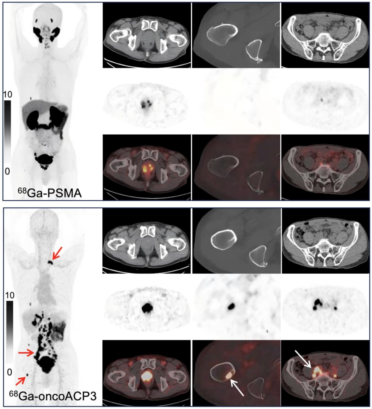
However, when all 318 lesions from the 65 patients were analyzed together, no significant difference in lesion-level uptake was observed between the two tracers (SUVmax 12.3 [IQR 5.4–20.8] versus 11.0 [IQR 5.8–17.7], p = 0.118). Regarding lesion detection, 68Ga-OncoACP3 identified 5 additional primary tumors, 1 lung metastasis, 16 lymph-node metastases, and 3 bone metastases compared with 68Ga-PSMA-11, but missed 1 liver metastasis, 4 lymph-node metastases, and 4 bone metastases. Importantly, due to nonspecific skeletal uptake, 68Ga-PSMA-11 generated 20 false-positive bone lesions, whereas 68Ga-OncoACP3 showed no false-positive bone lesions. 11 patients who underwent dual-time-point 68Ga-OncoACP3 PET, 69 lesions were analyzed. Delayed imaging at 2 hours demonstrated significantly higher tumor uptake than at 1 hour (SUVmax 13.6 [IQR 4.0–25.4] versus 12.0 [IQR 3.5–20.3], p < 0.001), indicating progressive tumor accumulation over time.
Liang Zhao concluded this presentation discussing a head-to-head comparison of 68Ga-OncoACP3 and 68Ga-PSMA-11 PET for imaging prostate cancer with the following take-home points:
- 68Ga-OncoACP3 demonstrated significantly higher uptake than 68Ga-PSMA-11 in primary prostate tumors and metastatic lymph nodes in patients with treatment-refractory disease, detected more primary tumors, and showed no false-positive bone lesions, while overall lesion-level uptake was comparable between the two tracers
- These findings indicate that 68Ga-OncoACP3 can serve as a valuable complementary imaging agent to 68Ga-PSMA-11, particularly in patients with heterogeneous or low PSMA expression
- Moreover, the markedly lower background uptake in most normal organs and the progressive increase in tumor uptake on delayed imaging suggest that OncoACP3 is a highly promising platform for labeling with therapeutic radionuclides such as 177Lu or 225Ac for targeted radioligand therapy in prostate cancer
References:
Presented by: Liang Zhao, MD, PhD, The First Affiliated Hospital of Xiamen University
Written by: Zachary Klaassen, MD, MSc – Urologic Oncologist, Associate Professor of Urology, Georgia Cancer Center, Wellstar MCG Health, @zklaassen_md on Twitter during the Society of Nuclear Medicine and Molecular Imaging (SNMMI) 2026 Annual Meeting, Los Angeles, CA, Sat, May 30 – Tues, Jun 2, 2026.