(UroToday.com) The 2025 Society of Nuclear Medicine and Molecular Imaging (SNMMI) Annual Meeting held in New Orleans, LA, was host to an Oncology Discovery and Translational session. Dr. Philipp Backhaus presented the first results of a study of prostatic acid phosphatase (ACP3) imaging with [68Ga]Ga-OncoACP3-DOTA PET in prostate cancer patients.
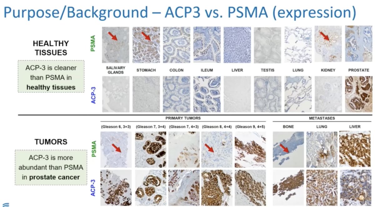
What makes ACP3 an attractive theranostic target? ACP3 is more abundant than PSMA in prostate cancer and is less commonly expressed in healthy tissues, compared to PSMA.
Using data from their DNA-encoded small molecule library, Philochem AG developed OncoACP3-DOTA, a high affinity ligand.

This ligand was evaluated in small animal experiments using microPET, with the production and use of [68Ga]Ga-OncoACP3-DOTA subsequently permitted under section 13.2b of German Pharmaceutical Law (AMG). This study, presented by Dr. Backhaus, was a retrospective analysis of the 1st 25 patients scanned with [68Ga]Ga-OncoACP3-DOTA and had available PSMA scans. In this analysis, the study investigators evaluated the biodistribution, tumor uptake, and diagnostic performance of this radiotracer.
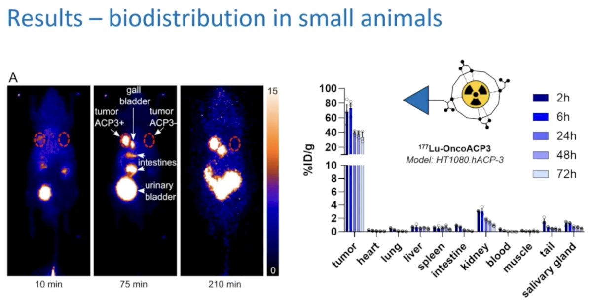
Illustrated below are the PET images with [68Ga]Ga-OncoACP3-DOTA in mouse models that had ACP-3 positive and negative tumors implanted in the left and right shoulders, respectively. We can clearly see intense uptake in the ACP3+ tumors and no uptake in the ACP3- tumors.
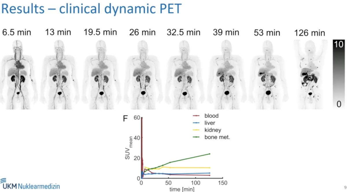
This study included 25 prostate cancer patients with available [68Ga]Ga-OncoACP3-DOTA-PET results. Of these 25 patients, 23 had [18F]PSMA-1007 PETs available for comparison of biodistribution. The diagnostic performance of [68Ga]Ga-OncoACP3-DOTA-PET was compared to that of [18F]PSMA-1007 and [68Ga]Ga-PSMA-11 using 27 matched PET scans. Significantly lower amounts of the [68Ga]Ga-OncoACP3-DOTA tracer were injected (146 +/- 38 MBq), compared to the PSMA radiotracer (233 +/- 51 MBq), and these patients were scanned at earlier timepoints (62 +/- 33 minutes versus 109 +/-21 minutes). Patients were scanned using a Siemens Biograph mCT or mMR, some dynamically. Of the 25 imaged patients, 7 had evidence of biochemical recurrence, and 18 had known metastatic disease. There were no side effects with PET imaging.
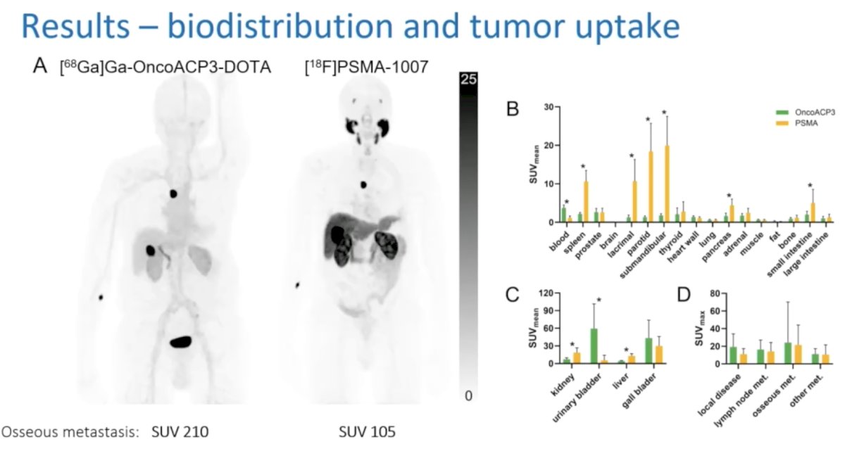
In a head-to-head comparison of [18F]PSMA-1007 PET and [68Ga]Ga-OncoACP3-DOTA-PET in one patient, Dr. Backhaus noted that the biodistribution is much cleaner with [68Ga]Ga-OncoACP3-DOTA, with no salivary or lacrimal gland uptake and much lower uptake in the liver, small intestine, spleen, and pancreas. The radiotracer uptake in the bone metastases was much higher with the [68Ga]Ga-OncoACP3-DOTA PET (210 versus 105) in this patient, but Dr. Backhaus noted that in the overall cohort, there were no significant differences in SUV expression between the two PET imaging modalities.
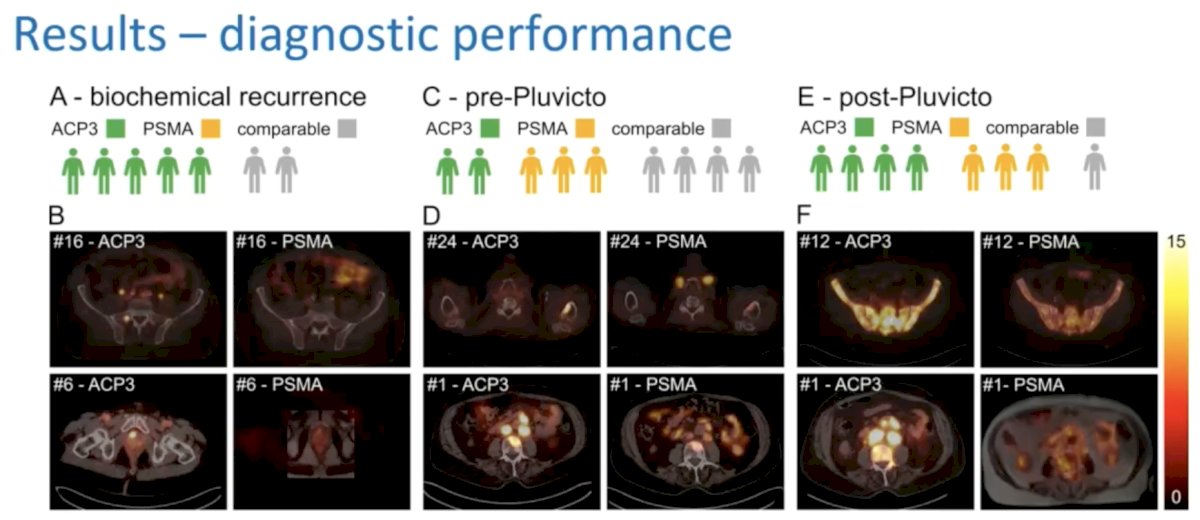
Overall, complementary uptake patterns were observed for the two radiotracers (i.e., some patients had ACP3+/PSMA- lesions and others ACP3-/PSMA+ lesions), but overall, OncoACP3 had superior performance (tumor uptake and tumor-to-organ ratio). There was no association between OncoACP3 uptake and Gleason Score or number of prior lines of therapy.

OncoACP3 triggered changes in the diagnostic work-up in 7/25 patients and changes in the therapeutic management plans in 9/25.
Limitations to this study include:
- Clinical reasoning for PET likely induced a bias towards low PSMA (and ACP3) expression
- The performance of ACP3 PET 16 days later, on average, after the PSMA PET may have biased results towards improved detection with ACP3 PET
- Small cohort of patients (n=25)
- Histologic verification of lesions was rarely performed
Dr. Backhaus concluded as follows:
- ACP3 is a highly promising theranostic target
- OncoACP3-DOTA shows potential to complement PSMA PET in low PSMA expressing cancers
Presented by: Philipp Backhaus, University of Münster, EIMI; University Hospital Münster, Munster, Germany
Written by: Rashid K. Sayyid, MD, MSc – Robotic Urologic Oncology Fellow at The University of Southern California, @rksayyid on Twitter during the 2025 Society of Nuclear Medicine and Molecular Imaging (SNMMI) Annual Meeting, New Orleans, LA, June 21st – 24th, 2025