(UroToday.com) The 2025 SNMMI annual meeting featured a prostate cancer and molecular imaging session and a presentation by Dr. Ida Sonni discussing a prospective, randomized trial comparing PSMA PET/CT and MRI-guided versus MRI only-guided prostate biopsy for identification of clinically significant prostate cancer. MRI is a well-established tool for prostate biopsy guidance, improving detection of clinically significant prostate cancer compared to systematic TRUS–guided biopsy. However, MRI has limitations, as it may miss smaller, less aggressive clinically significant prostate cancer that may potentially be identified by PSMA PET/CT. PSMA PET has transformed prostate cancer management, but its pre-biopsy role remains under investigation. This study assessed whether 18F-DCFPyL PSMA PET/CT fused with mpMRI improves clinically significant prostate cancer detection compared to mpMRI alone in men with PI-RADS 4 lesions.
This single-center, randomized phase II trial conducted at the Greater Los Angeles VA enrolled men with at least one PI-RADS 4 lesion on mpMRI. Exclusion criteria include the presence of PI-RADS 5 lesions or an interval between MRI and enrollment exceeding 9 months. Participants were randomly assigned in a 1:1 ratio to undergo MRI-only TRUS biopsy (Arm 1) or PSMA PET/CT + mpMRI-guided biopsy (Arm 2). In both arms, a 12-core systematic biopsy was performed as part of standard of care:

Lesions were contoured on MRI (PI-RADS 4, 3) and PET/CT (PRIMARY 3, 4, 5) by experienced physicians, and fused PET/MRI images were obtained to guide the biopsy. The primary outcome was the positive predictive value for detecting clinically significant prostate cancer at both lesion and patient levels. The secondary outcome was the positive predictive value for all prostate cancer detection (clinically significant and non-significant) in Arm 1 versus Arm 2. Dr. Sonni and colleagues further investigated the positive predictive value for clinically significant prostate cancer detection on PSMA PET/CT versus mpMRI within Arm 2, as well as the association between PET quantitative measures (ie. SUVmax and SUVmean) and the detection of clinically significant prostate cancer.
The flow chart below demonstrates patient selection to date for this trial:

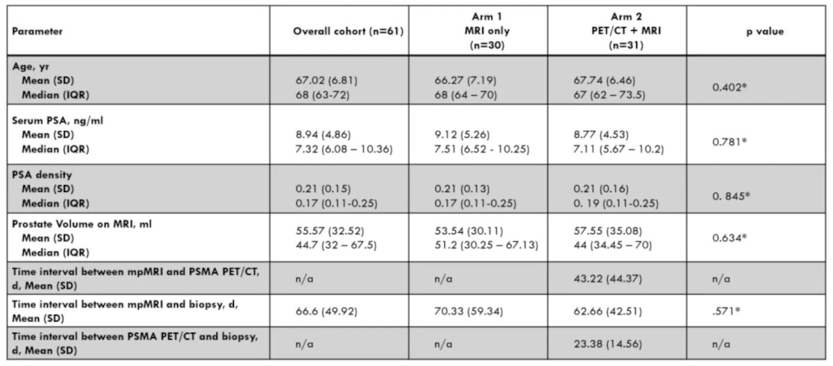
Preliminary results from the first 61 participants randomized for the trial showed no statistically significant differences in baseline characteristics (age, PSA, PSA density, prostate volume):
At the lesion level, clinically significant prostate cancer positive predictive value was 62% in Arm 1 and 61% in Arm 2 (p = 0.914), while the all prostate cancer positive predictive value was 72% and 75% (p = 0.932), respectively. At the patient level, clinically significant prostate cancer positive predictive value was 73% in Arm 1 versus 77% in Arm 2 (p = 0.71), and the all prostate cancer positive predictive value was 80% versus 84% (p = 0.69), respectively.
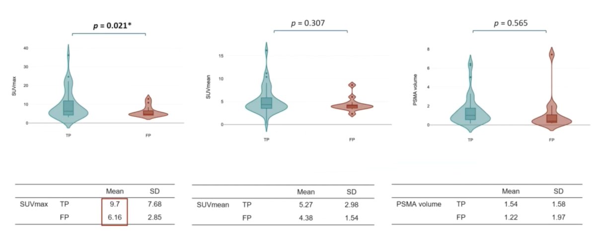
In Arm 2, PET/CT demonstrated a clinically significant prostate cancer positive predictive value of 78% versus 77% for MRI at the patient level (p = 0.233), and 74% for PET/CT versus 59% for MRI at the lesion level (p = 0.006). For all prostate cancer detection, the positive predictive value at the patient level was 85% for PET/CT versus 81% for MRI (p = 0.214), and at the lesion level was 80% for PET/CT versus 71% for MRI (p = 0.012). SUVmax was significantly higher in true positives versus false positives (p = 0.021), whereas SUVmean (p = 0.307), and PSMA volume (p = 0.565) had no association:
Dr. Sonni concluded her presentation discussing a prospective, randomized trial comparing PSMA PET/CT and MRI-guided versus MRI only-guided prostate biopsy for identification of clinically significant prostate cancer with the following take home points:
- At the patient level, there is a trend showing better detection of clinically significant prostate cancer for patients undergoing prostate biopsy using the combined 18F-DCFPyL PSMA PET/CT and mpMRI for biopsy guidance compared to mpMRI alone, but this does not reach statistical significance
- At the lesion level, combined PSMA PET/CT and MRI obtained similar positive predictive value to mpMRI alone
- PSMA PET/CT identified more clinically significant prostate cancer compared to mpMRI, but this difference was not significant at the patient level
- Further analysis will be reported upon the study's completion
Presented by: Ida Sonni, MD, UCLA, Los Angeles, CA
Written by: Zachary Klaassen, MD, MSc – Urologic Oncologist, Associate Professor of Urology, Georgia Cancer Center, Wellstar MCG Health, @zklaassen_md on Twitter during the 2025 Society of Nuclear Medicine and Molecular Imaging (SNMMI) Annual Meeting, New Orleans, LA, June 21st – 24th, 2025