(UroToday.com) The 2025 Society of Nuclear Medicine and Molecular Imaging (SNMMI) Annual Meeting held in New Orleans, LA, was host to an Oncology Discovery and Translational session. Dr. Gary Ulaner presented a phase I trial of 61Cu-NODAGA-PSMA I&T for prostate cancer patients.
Currently, there are numerous PSMA-targeted agents available for use as radioisotopes for prostate PET imaging, with 68Ga-PSMA-11, 18F-Piflulolastat (Pylarify), and 18F-Flotufolastat (Posluma) approved by the FDA in this setting.

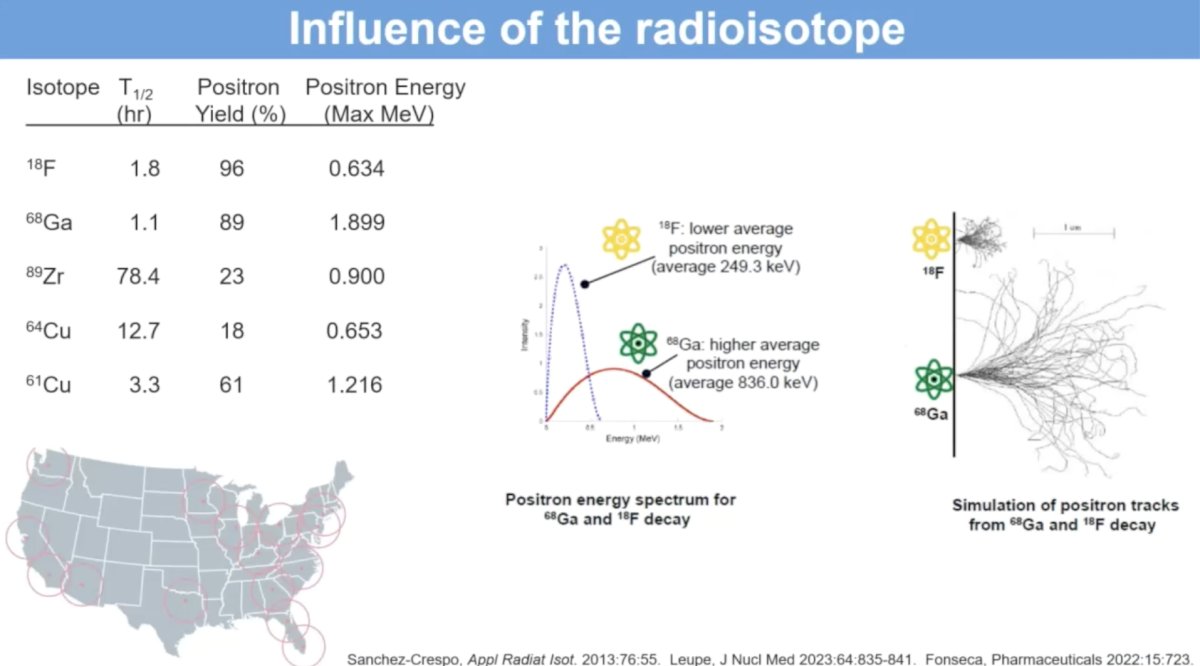
Dr. Ulaner argued that 18F remains the ‘gold standard’ radioisotope in this setting, with the highest positron yield (96%) and a low positron energy (0.634 Max MeV), which are characteristics that favor high resolution imaging.
61Cu is a relatively newer radioisotope with a relatively high positron yield (61%) and a long half-life (3.3 hours), which is advantageous for delayed imaging and allows for long-distance shipping.
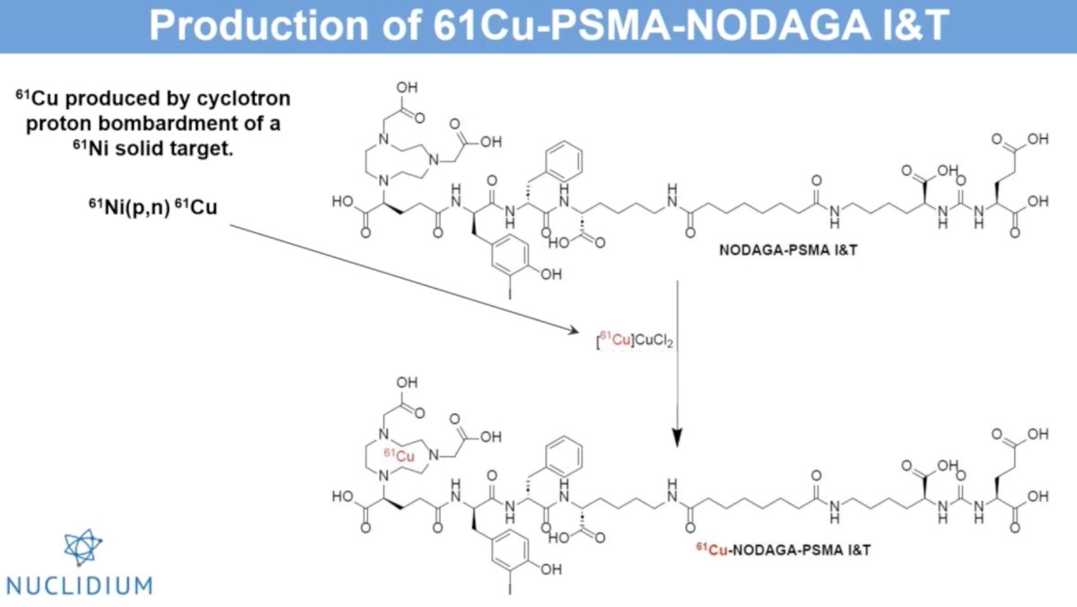
61Cu is produced by cyclotron proton bombardment of a 61Ni solid target and is being chelated with NODAGA-PSMA I&T.
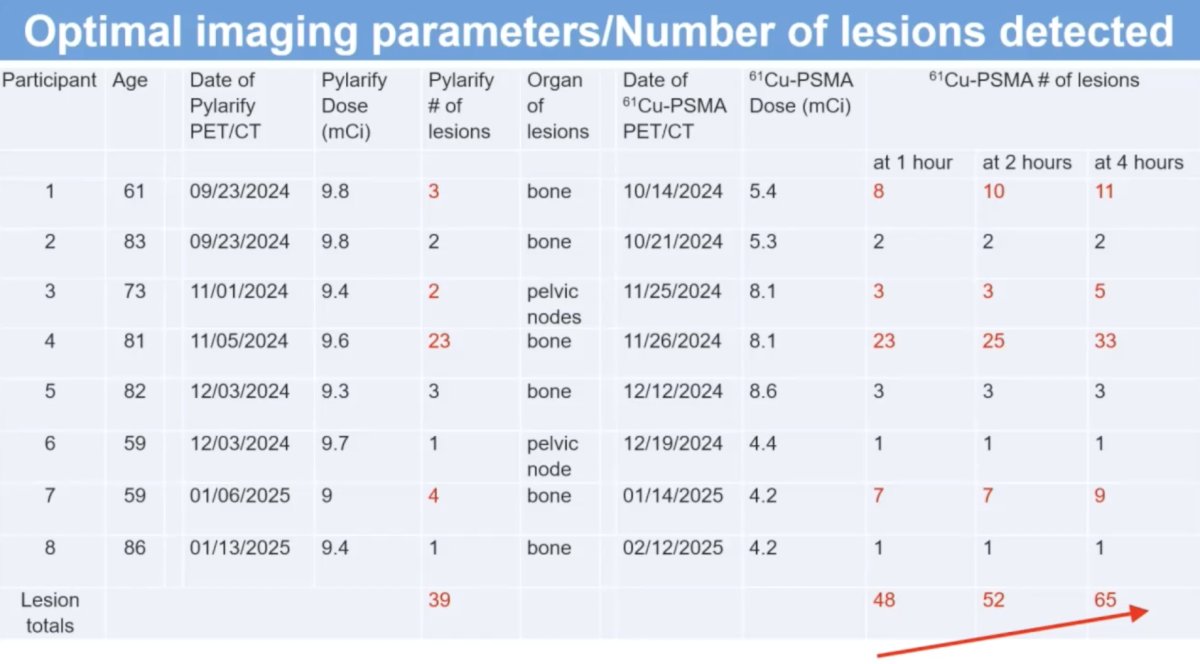
This is a phase I trial that aimed to evaluate the safety, dosimetry, and optimal imaging parameters of 61Cu-NODAGA-PSMA I&T. It included patients with PSMA-avid disease on Pylarify PET/CT. The 61Cu-NODAGA-PSMA I&T was produced at Pharmacologic Los Angeles and ground transported to Hoag. Eligible patients received IV 61Cu-NODAGA-PSMA I&T at doses of 4.2-8.6 mCi with PET scans performed at 1, 2, and 4 hours. Blood samples, vitals, and adverse reactions were assessed at set intervals, as detailed below.

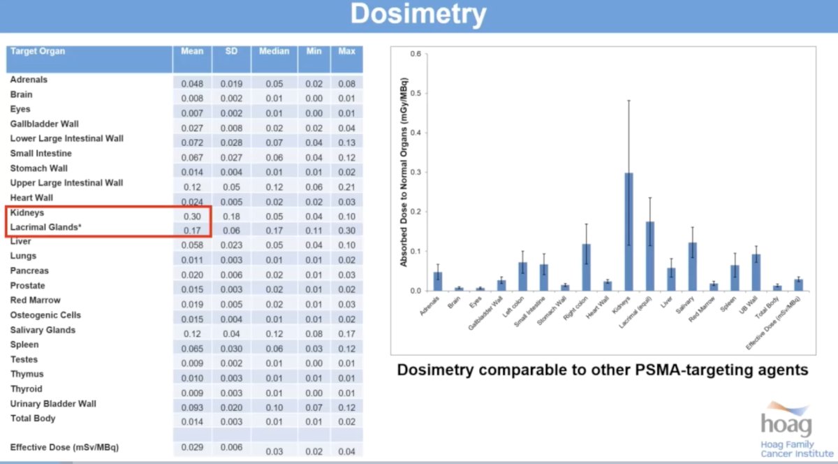
No side effects were identified. From a dosimetry standpoint, the two most critical organs are the kidneys and the lacrimal glands. The total absorbed dose in the kidneys was only 0.29 (mGy/MBq), which is comparable to other PSMA PET imaging agents.
From an efficacy standpoint, all lesions detected on Pylarify PET were detected on 61Cu PSMA PET. In 4/8 patients, the same number of lesions were detected on both PET scans. In the remaining 4 patients, more lesions were actually detected on the 61Cu PSMA scan. As summarized in the lowest row, a higher total number of lesions were detected on 61Cu PSMA PET (48, 52, and 65 at 1, 2, and 4 hours, respectively versus 39 lesions on Pylarify PET).
Why do we see more lesions on 61Cu PSMA PET, and why does this yield increase over time from 1 to 4 hours? This is related to the SUV of the lesions and the background noise. As seen below, the mean lesional SUVmax increases from 23.3 at 1-hour post-61Cu administration to 30.4 and 33.2 at 2- and 4-hours post-administration. Conversely, the background liver and bone SUVmax correspondingly decrease.

Important limitations to this study include the lack of histology – this precludes commenting on the sensitivity and specificity of this imaging modality in this study. There was a limited number of subjects. All PET scans were interpreted by a single reader.
Dr. Ulaner concluded his presentation of this phase I trial of 61Cu-NODAGA-PSMA I&T PET/CT as follows:
- 61Cu has favorable physical characteristics, including positron yield, positron energy, and a 3.3-hour half-life allowed for 4-hour delayed imaging
- There were no adverse events and favorable dosimetry
- As 61Cu-PSMA uptake time increases, lesional SUV increases, while background SUV decreases
- As 61Cu-PSMA uptake time increases, the number of lesions detected also increases:
- 1-hour 61Cu-PSMA PET/CT detects more lesions than Pylarify PET/CT
- 4-hour 61Cu-PSMA PET/CT detects 60% more lesions than Pylarify PET/CT
- These results support the development of 61Cu-NODAGA-PSMA I&T as an imaging agent for patients with prostate cancer. More broadly, this opens new opportunities for the construction of novel 61Cu-labeled PET radiotracers.
Presented by: Gary Ulaner, MD, PhD, Clinical Professor of Radiology and Cancer Biology, Hoag Family Cancer Institute, University of Southern California, Los Angeles, CA
Written by: Rashid K. Sayyid, MD, MSc – Robotic Urologic Oncology Fellow at The University of Southern California, @rksayyid on Twitter during the 2025 Society of Nuclear Medicine and Molecular Imaging (SNMMI) Annual Meeting, New Orleans, LA, June 21st – 24th, 2025
Related content: Early-Phase Study Investigates Copper-61 PSMA PET for Prostate Cancer Imaging - Gary Ulaner