(UroToday.com) The 2025 European Society of Medical Oncology (ESMO) Annual Congress, held in Berlin, Germany between October 17th and 21st was host to the session Mini Oral session 2: GU tumours, renal & urothelial Dr. Rana R. McKay presented abstract 2593MO - Lymphocyte activation gene-3 (LAG3) expression patterns and immunotherapy (IO) response in metastatic renal cell carcinoma (mRCC).
Dr. McKay began by highlighting that immunotherapy combinations have become the new frontline standard of care in renal cell carcinoma (RCC). However, treatment outcomes remain highly heterogeneous, underscoring the need for reliable biomarkers. Although a wide range of biomarkers have been explored in RCC, none are currently used in clinical practice. LAG-3, an inhibitory receptor on T-cells that suppresses activation and promotes exhaustion, has emerged as a potential marker. Its expression alone or in combination with PD-1/PD-L1 and other immune exhaustion markers has been linked to an inflamed tumor microenvironment (TME). The objective of this study was to investigate LAG-3 RNA expression as a potential biomarker in patients with RCC receiving frontline immunotherapy.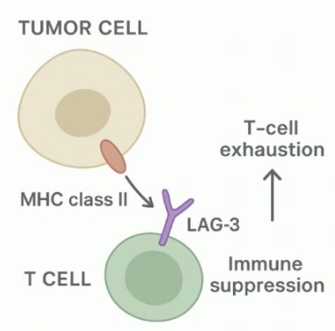
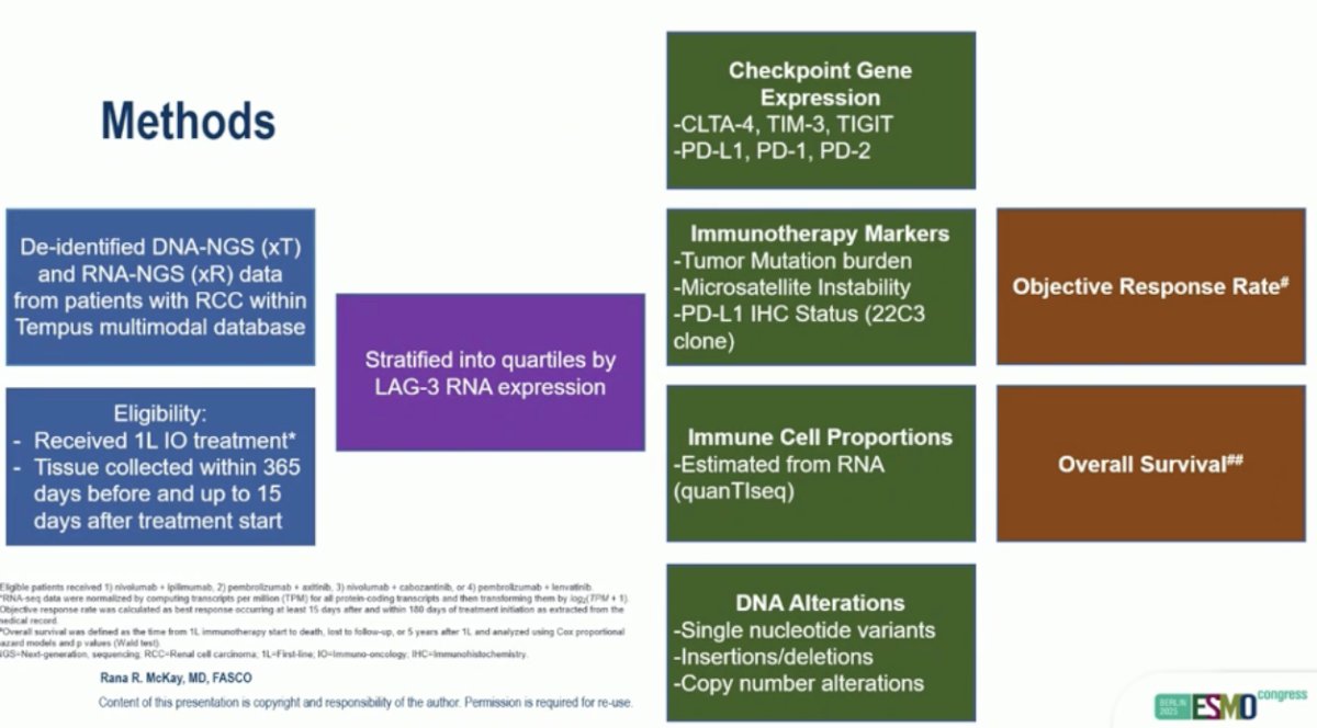
In this analysis, they used de-identified DNA-NGS (xT) and RNA-NGS (xR) data from patients with RCC available in the Tempus multimodal database. Eligible patients had received first-line (1L) immunotherapy and had tissue collected within 365 days before or up to 15 days after treatment initiation. Patients were stratified into quartiles based on LAG-3 RNA expression. The study assessed checkpoint gene expression (including CTLA-4, TIM-3, TIGIT, PD-1, and PD-L1), immune cell proportions estimated from RNA, immunotherapy biomarkers such as tumor mutation burden and microsatellite instability, and DNA alterations.
The primary outcomes of the study were objective response rate (ORR) and overall survival (OS), evaluated according to levels of LAG-3 expression. Methods are outlined below.
A total of 566 patients were included in this analysis. Baseline characteristics were generally well balanced across LAG-3 RNA expression quartiles. However, liver metastases were more frequent among patients in the highest quartile of LAG-3 expression (Q4: 17%) compared with lower quartiles (Q1–Q3: 13%, 9%, and 13%, respectively; p=0.019)

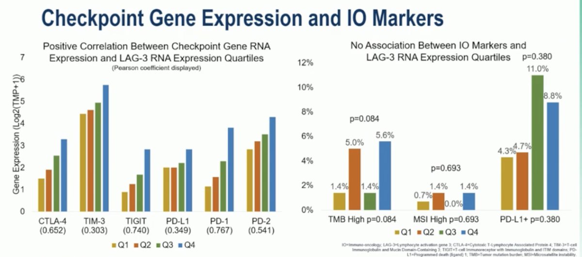
Checkpoint gene expression analysis showed a positive correlation between LAG-3 RNA expression and other immune checkpoint genes, including CTLA-4, TIM-3, TIGIT, PD-1, and PD-2. These findings suggest that higher LAG-3 expression is associated with a more inflamed tumor microenvironment.
Conversely, there was no significant association between LAG-3 RNA expression and other immunotherapy biomarkers such as tumor mutation burden (TMB), microsatellite instability (MSI), or PD-L1 positivity, indicating that LAG-3 may serve as an independent marker of immune activation.
Moreover, they found a positive association between immune cell fraction and LAG-3 RNA expression quartiles. Higher LAG-3 expression correlated with increased infiltration of multiple immune cell populations, including B cells, M1 and M2 macrophages, NK cells, and CD8+ T cells (all p<0.001). This pattern suggests that tumors with elevated LAG-3 RNA expression exhibit an inflamed yet exhausted immune profile, characterized by abundant immune infiltration alongside signs of immune suppression and exhaustion.

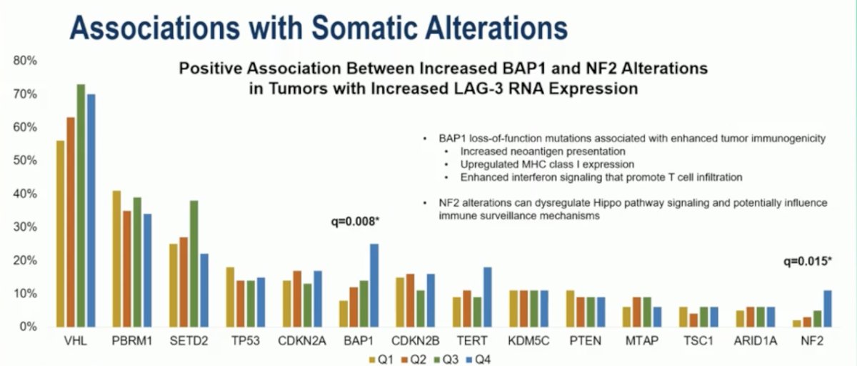
Dr. McKay highlighted that tumors with higher LAG-3 RNA expression showed a positive association with somatic alterations in BAP1 (q=0.008) and NF2 (q=0.015). They noted that BAP1 loss-of-function mutations are linked to enhanced tumor immunogenicity through increased neoantigen presentation, upregulated MHC class I expression, and augmented interferon signaling that promotes T-cell infiltration. Similarly, NF2 alterations can dysregulate Hippo pathway signaling, potentially impairing immune surveillance mechanisms. Together, these findings suggest that increased LAG-3 expression may coexist with genetic changes that contribute to an inflamed but immunologically exhausted tumor microenvironment.
Higher LAG-3 RNA expression was associated with improved objective response rates and a lower incidence of progressive disease (p=0.034). Patients in the highest quartile of LAG-3 expression (Q4) had the greatest proportion of partial responses (51%) and the lowest rates of progression (28%).

Notably, they found no significant association between LAG-3 RNA expression and overall survival. Median OS ranged from 25.7 months in the lowest quartile (Q1) to 35 months in the highest quartile (Q4), with no statistically significant differences observed across groups (HR 0.74; 95% CI, 0.46–1.18; p=0.2). These results suggest that while elevated LAG-3 expression correlates with increased immune activity and response, it does not independently predict long-term survival outcomes in patients receiving frontline immunotherapy for RCC.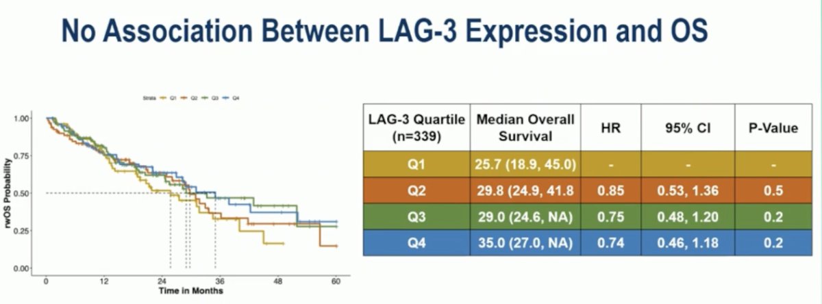
Dr. McKay concluded her presentation with the following key takeaways:
- Tumors with higher LAG-3 expression demonstrated a decreased proportion of liver metastases.
- LAG-3 RNA expression positively correlated with other checkpoint genes, suggesting severe T-cell dysfunction and adaptive immune resistance.
- Tumors with high LAG-3 expression exhibited a complex immunological phenotype, marked by robust effector cell infiltration (CD8+ T cells, NK cells, M1 macrophages, B cells) alongside concurrent immunosuppressive mechanisms (M2 macrophages, Tregs), indicating an adaptive resistance state.
- High LAG-3 expression was associated with increased BAP1 and NF2 alterations, both linked to enhanced tumor immunogenicity.
- Greater responses were observed among tumors with increased LAG-3 expression; however, overall survival remained similar across LAG-3 RNA expression quartiles.
- Further data and protein-level correlation are needed to validate these findings
Presented by: Rana R. McKay, MD, Medical Oncologist, Clinical Professor of Medicine, UC San Diego School of Medicine, La Jolla, United States of America.
Written by: Julian Chavarriaga, MD – Urologic Oncologist at Cancer Treatment and Research Center (CTIC) via Society of Urologic Oncology (SUO) Fellow at The University of Toronto. @chavarriagaj on Twitter during the 2025 European Society of Medical Oncology (ESMO) Annual Congress held in Berlin, Germany, between October 17th and 21st.