(UroToday.com) The 2025 European Association of Urology (EAU) Annual Meeting held in Madrid, Spain was host to the Biomarkers to guide peri-operative management in Uro-oncology Plenary Session. Dr. Valeria Panebianco delivered a presentation on imaging as potential biomarkers to guide peri-operative management in urologic oncology.
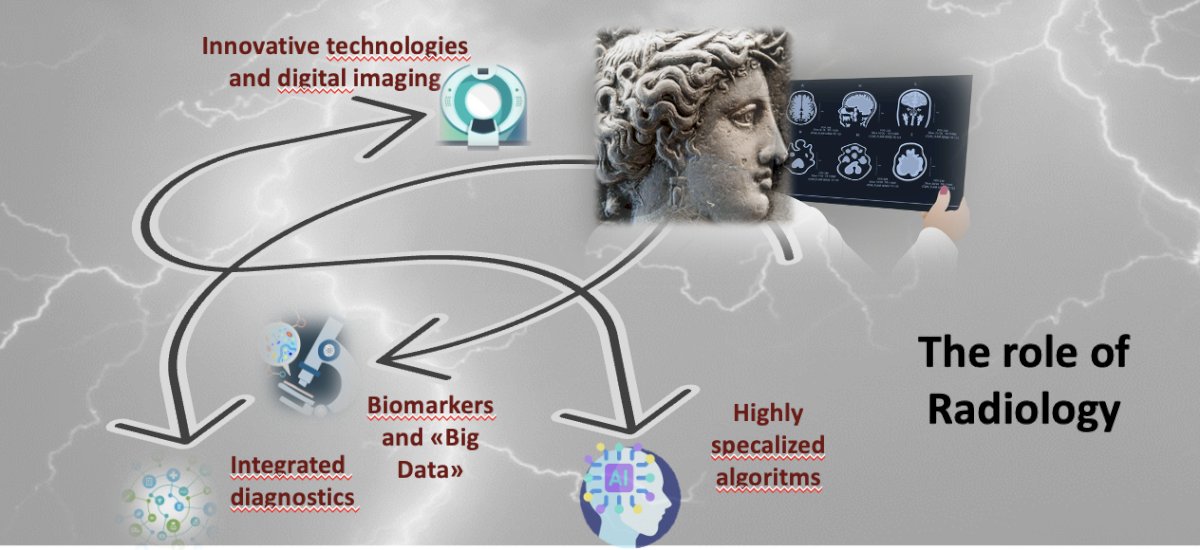
Nowadays, patients navigate different pathways when seeking medical care, including the clinical pathway, precision diagnostic pathway, computational pathway, and digital & virtual pathway. Dr. Panebianco emphasized that there is no precision treatment without precision diagnostics. The challenge lies in identifying the specific diagnostic tool for each clinical question and selecting the right biomarker. Currently, we are experiencing the "perfect clinical storm," where innovative technologies, digital imaging, integrated diagnostics, biomarkers, big data, and highly specialized algorithms converge. This raises an important question: what is the role of radiology in this evolving landscape?
An imaging biomarker is a biological feature detectable in an image. Imaging biomarkers (IBs) are integral to the routine management of patients with cancer, with key applications including detection and screening, staging and grading, and monitoring treatment response. Examples of imaging biomarkers include:
- Organ and lesion volume
- TNM staging (CT, MRI, PET)
- ACR-RADS
- SUVmax in PET scans
- Ktrans in MRI for vascular permeability
- ADC in diffusion MRI for cellularity changes
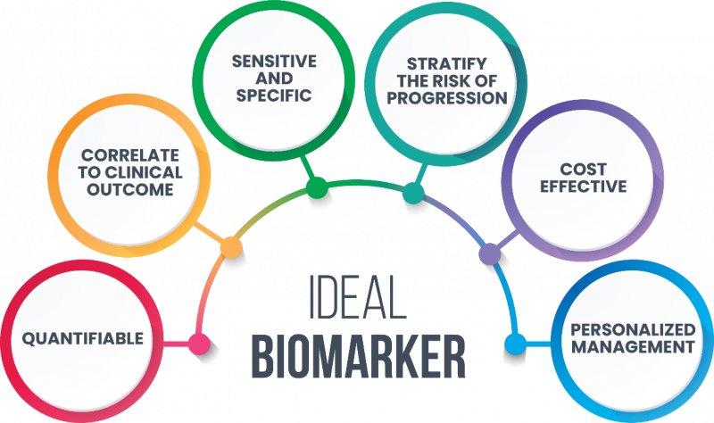
A biomarker definition according to the FDA and EMA requires biomarkers to be quantifiable measures of a specific disease or response to treatment, with predictive biomarkers being particularly relevant. The ideal biomarker should be quantifiable, sensitive, specific, cost-effective, predictive, and correlate with clinical outcomes.
In Europe, interest has grown in Imaging repositories. There are many recent projects, however, she highlighted five research projects, developing digital storage systems for oncological imaging data, with specific attention to Artificial Intelligence. These projects lie on the Horizon 2020

Imaging biomarkers in Europe are regulated by EIBALL (European Imaging Biomarkers Alliance) under the ESR, while in the USA, they fall under QIBA (Quantitative Imaging Biomarkers Alliance) from the RSNA. These organizations play a critical role in advancing the standardization and clinical adoption of imaging biomarkers, ensuring their reliability and effectiveness in both research and patient care.
A key objective is to establish a functional and up-to-date biomarker profile through the development of a digital biomarker repository. This repository would serve as a comprehensive reference, integrating validated biomarkers across different imaging modalities to enhance diagnostic precision and treatment decision-making.
Another essential goal is the definition of imaging biomarkers for both oncological and non-oncological diseases. By categorizing and validating biomarkers specific to different pathologies, clinicians can improve disease characterization, risk stratification, and therapeutic monitoring.
Finally, enabling and encouraging the clinical use of biomarkers requires setting rigorous standards for data acquisition and image processing. Standardization ensures consistency in biomarker interpretation across institutions, facilitating their integration into routine clinical workflows and improving patient outcomes.
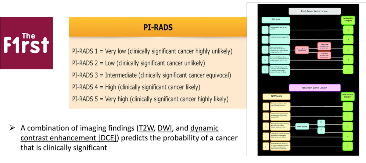
Prostate cancerDr. Panebianco discussed the role of scoring systems and RADS (Reporting and Data Systems) in prostate MRI assessment, PI-RADS was the first to use a combination of imaging findings—T2-weighted (T2W) imaging, diffusion-weighted imaging (DWI), and dynamic contrast enhancement (DCE) to help predict the likelihood of clinically significant prostate cancer.
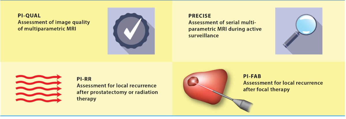
Multiparametric MRI (mpMRI) is increasingly being used beyond its initial role in prostate cancer detection, expanding into areas such as active surveillance and the evaluation of local recurrence after prostatectomy, radiation therapy, or focal therapy. To enhance the consistency and accuracy of MRI interpretation in these settings, new prostate MRI scoring systems are being developed. These include PI-QUAL for imaging quality assessment of MRI, PRECISSE for active surveillance, PI-RR for evaluating recurrence after prostatectomy or radiation therapy, and PI-FAB for assessing post-focal therapy changes.1
Similar initiatives are emerging in the United States, with many being supported by the National Institutes of Health (NIH). Key efforts include resources like the Cancer Imaging Archive and the Imaging Data Commons, which facilitate the collection, standardization, and analysis of imaging data to improve clinical decision-making and research in prostate cancer management.
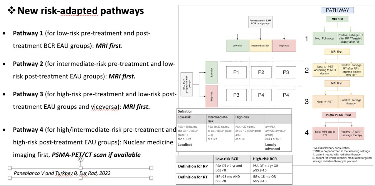
The Prostate Imaging for Recurrence Reporting (PI-RR) scoring system has been validated as an imaging biomarker for detecting prostate cancer recurrence after both radical prostatectomy and radiation therapy, the PI-RR has an assessment score that goes from one to five. In a single-center retrospective observational study of 100 patients, PI-RR demonstrated excellent inter-reader agreement across four readers, with an intraclass correlation coefficient (ICC) of 0.87. Specifically, for patients who underwent radiation therapy (n = 48), the area under the receiver operating characteristic curve (AUC) of PI-RR ranged from 0.77 to 0.92 across readers. For those who had radical prostatectomy (n = 52), the AUC ranged from 0.80 to 0.88, indicating strong performance in detecting local recurrence.2
Based on this Dr. Panebianco et al recommended a new risk-adapted pathway for patients with biochemical recurrence has been proposed, emphasizing the role of MRI as the first imaging modality. This approach stratifies patients into four distinct pathways based on clinical and imaging findings, optimizing the detection of local versus distant recurrence and guiding subsequent management decisions. The proposed pathway highlights the increasing role of MRI in the early identification of recurrence, potentially improving treatment planning and patient outcomes.3
The PRECISE score has been validated as an imaging biomarker in active surveillance for prostate cancer. This scoring system evaluates various MRI features, including lesion conspicuity and size, to assess disease progression over time. PRECISE assigns a score ranging from 1 to 5, where:
- 1 or 2 indicates radiological regression.
- 3 reflects radiological stability.
- 4 or 5 denotes radiological progression.
By incorporating these criteria, the PRECISE score helps guide clinical decision-making, ensuring timely intervention for patients who exhibit signs of disease progression.4

Below is a summary algorithm to guide the selection of the appropriate prostate MRI scoring system based on different clinical scenarios:

In bladder cancer, MRI can serve as a biomarker for staging, with VI-RADS (Vesical Imaging-Reporting and Data System) being a widely used scoring system. VI-RADS classifies patients based on the likelihood of muscle-invasive bladder cancer (MIBC) and helps guide treatment decisions both before and after TURBT (transurethral resection of bladder tumor). By providing a standardized assessment of tumor invasion depth, VI-RADS improves clinical decision-making and may reduce unnecessary overtreatment or delays in appropriate therapy.

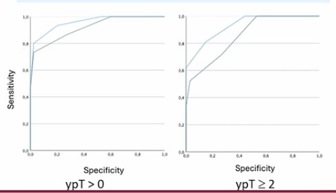
More recently, MRI has been proposed as a biomarker for assessing response to neoadjuvant therapy in bladder cancer. This risk-scoring system, known as nac-VI-RADS, aims to evaluate radiological complete response, defined as no residual disease detectable on follow-up imaging after four cycles of neoadjuvant chemotherapy (NAC).

In a study of 57 patients who received three to four cycles of cisplatin-based NAC, inter-reader agreement was assessed, showing an optimal agreement with a kappa (K) value of 0.85. This suggests that nac-VI-RADS could serve as a reliable tool for evaluating treatment response (ypT0) and guiding subsequent management decisions.5
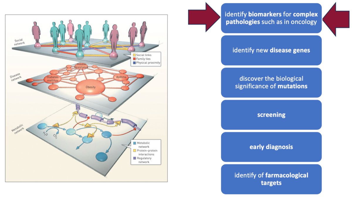
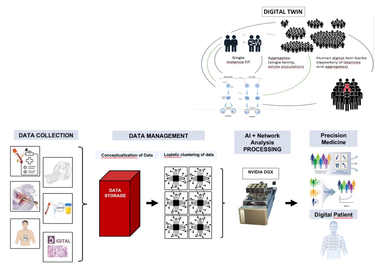
The computational pathway in medical imaging and diagnostics involves both network analysis and artificial intelligence (AI). Network analysis plays a crucial role in integrating diverse data sources, identifying underlying causes, and defining aggregation clusters with shared characteristics. This approach enhances the understanding of complex disease patterns and facilitates more precise patient stratification. Meanwhile, AI contributes by standardizing data acquisition, processing, and interpretation. Through advanced algorithms, AI can streamline image analysis, improve diagnostic accuracy, and support clinical decision-making, ultimately advancing precision medicine in oncology and beyond.
In oncology, network analysis (NA) is a powerful tool for exploring complex computational and clinical biomarkers, particularly for early detection and screening. But how does it work? NA facilitates data integration, enabling the recognition of underlying causes and the definition of aggregation clusters with similar characteristics. By analyzing vast datasets, network analysis can help identify:
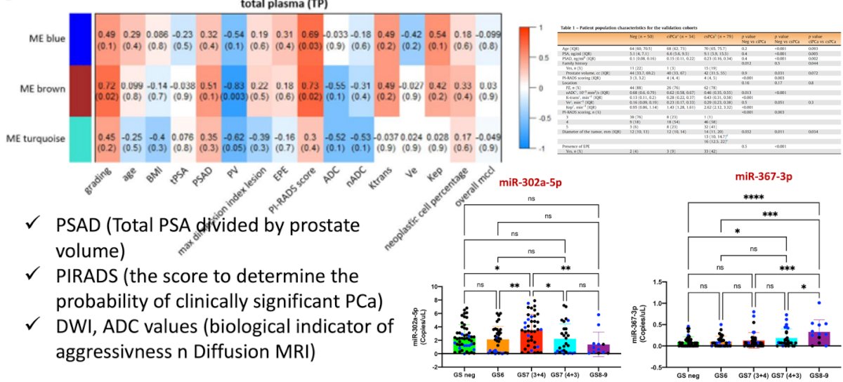
Dr. Panebianco highlighted that network analysis has been applied in prostate cancer. The first example involves pre-biopsy triage for patients at risk of prostate cancer (PCa). A proposed diagnostic paradigm shift integrates molecular data and MRI biomarkers as part of the pre-biopsy evaluation. NA could enhance early detection by integrating clinical data, miRNA expression profiles, and imaging biomarkers, helping to better stratify patients and reduce unnecessary biopsies.
Dr. Panebianco and colleagues conducted a prospective single-center cohort study involving 261 patients who underwent MRI, MRI-directed fusion biopsy, and circulating microRNA analysis. A network-based analysis was performed to identify MRI biomarkers and microRNA drivers associated with clinically significant prostate cancer. The study aimed to validate MRI biomarkers and circulating microRNAs as triage tests for patients undergoing prostate biopsy and to compare different diagnostic pathways to assess their impact on patient outcomes, particularly in terms of reducing unnecessary biopsies.
In this study, using network analysis, the investigators identified two microRNAs—miR-302 and miR-367—by integrating imaging data with clinical findings, both of which correlated with clinically significant prostate cancer. Key biomarkers included PSAD, PI-RADS, ADC values, and these circulating microRNAs. The findings suggest that combining MRI with novel circulating biomarkers from liquid biopsy can enhance risk stratification and improve the prediction of tumor aggressiveness.6
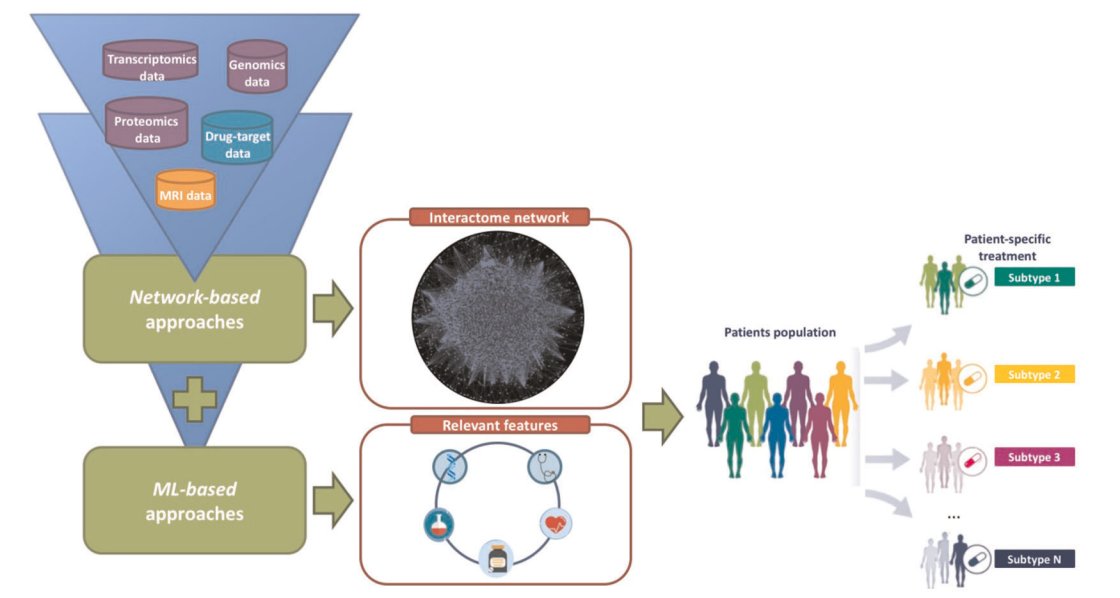
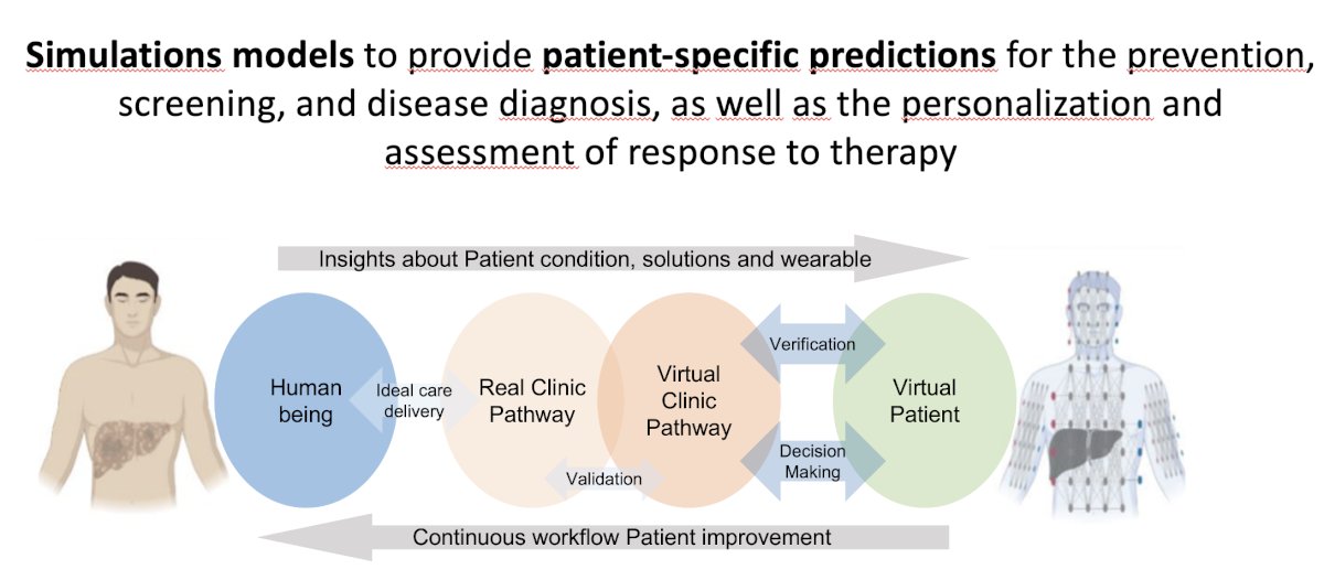
Virtual Pathway Future perspectives in radiology focus on the discovery of new disease-specific biomarkers, the use of computational profiles to implement advanced diagnostic and prevention models and the adoption of Digital Twin technology. The Digital Twin approach creates a dynamic model of an individual, integrating all relevant biological and clinical components to simulate disease progression and treatment responses, ultimately enhancing precision medicine strategies.
This is the proposed workflow between the virtual and the human patient:
Dr. Panebianco concluded her presentation with the following take home messages:
- The complexity and prognostic heterogeneity of oncological pathologies make it necessary to apply precision diagnostics
- Imaging morpho and functional pattern, RADS and scores can be considered as biomarkers
- Computational methods can be exploited to achieve the
- goals of precision medicine
- Network Analysis, AI, by integrating imaging data with omics data and other, is able to identify specific biomarkers for the disease
- Digital Twin can represent the definitive outcome, establishing a virtual pathway
Presented by: Valeria Panebianco, MD, Professor of Radiology - Director of Radiological Sciences, Oncology and Pathology Department at Sapienza University of Rome
Written by: Julian Chavarriaga, MD – Urologic Oncologist at Cancer Treatment and Research Center (CTIC) via Society of Urologic Oncology (SUO) Fellow at The University of Toronto. @chavarriagaj on Twitter during the European Association of Urology (EAU) 2025 Annual Meeting, Madrid, Spain, Fri, Mar 21 – Mon, Mar 24, 2025.
References:- Dias AB, Chang SD, Fennessy FM, Ghafoor S, Ghai S, Panebianco V, Purysko AS, Giganti F. New Prostate MRI Scoring Systems (PI-QUAL, PRECISE, PI-RR, and PI-FAB): AJR Expert Panel Narrative Review. AJR Am J Roentgenol. 2025 Feb;224(2):e2430956. doi: 10.2214/AJR.24.30956. Epub 2024 Apr 3. PMID: 38568038.
- Pecoraro M, Turkbey B, Purysko AS, Girometti R, Giannarini G, Villeirs G, Roberto M, Catalano C, Padhani AR, Barentsz JO, Panebianco V. Diagnostic Accuracy and Observer Agreement of the MRI Prostate Imaging for Recurrence Reporting Assessment Score. Radiology. 2022 Aug;304(2):342-350. doi: 10.1148/radiol.212252. Epub 2022 May 10. PMID: 35536130.
- Panebianco V, Villeirs G, Weinreb JC, Turkbey BI, Margolis DJ, Richenberg J, Schoots IG, Moore CM, Futterer J, Macura KJ, Oto A, Bittencourt LK, Haider MA, Salomon G, Tempany CM, Padhani AR, Barentsz JO. Prostate Magnetic Resonance Imaging for Local Recurrence Reporting (PI-RR): International Consensus -based Guidelines on Multiparametric Magnetic Resonance Imaging for Prostate Cancer Recurrence after Radiation Therapy and Radical Prostatectomy. Eur Urol Oncol. 2021 Dec;4(6):868-876. doi: 10.1016/j.euo.2021.01.003. Epub 2021 Feb 10. PMID: 33582104.
- Ponsiglione A, Brembilla G, Cuocolo R, Gutierrez P, Moreira AS, Pecoraro M, Zawaideh J, Barentsz J, Giganti F, Padhani AR, Panebianco V, Puech P, Villeirs G. ESR Essentials: using the right scoring system in prostate MRI-practice recommendations by ESUR. Eur Radiol. 2024 Nov;34(11):7481-7491. doi: 10.1007/s00330-024-10792-7. Epub 2024 May 23. PMID: 38780764; PMCID: PMC11519295.
- Dehghanpour A, Pecoraro M, Messina E, Laschena L, Borrelli A, Novelli S, Santini D, Simone G, Girometti R, Panebianco V. Diagnostic accuracy and inter-reader agreement of the nacVI-RADS for bladder cancer treated with neoadjuvant chemotherapy: a prospective validation study. Eur Radiol. 2024 Dec 31. doi: 10.1007/s00330-024-11327-w. Epub ahead of print. PMID: 39738561.
- Pecoraro M, Catanzaro G, Conte F, Besharat ZM, Messina E, Laschena L, Trocchianesi S, Splendiani E, Sciarra A, Catalano C, Paci P, Ferretti E, Panebianco V. Prospective Validation Study of a Novel Integrated Pathway Based on Clinical Features, Magnetic Resonance Imaging Biomarkers, and MicroRNAs for Early Detection of Prostate Cancer. Eur Urol Oncol. 2024 Feb;7(1):73-82. doi: 10.1016/j.euo.2023.05.008. Epub 2023 Jun 1. PMID: 37270379.