(UroToday.com) Gavin Hughes, a medical student at the University of Toronto, began his podium presentation with an anecdotal comparison between two patients with 100% calcium oxalate monohydrate (COM) stones where he found that the kidney stone in patient 1 was measured to have a higher Hounsfield units (HUs) of 1319 HUs than patient 2 with 1230 HUs. Furthermore, patient 1 was rated to have 3/10 fragmentation difficulty while patient 2 was rated 8/10 for fragmentation difficulty during percutaneous nephrolithotomy (PCNL) by the operating surgeon (Figure 1).
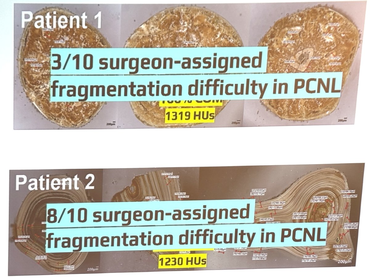
Figure 1. Example of two patients with 100% COM stones with measured HUs and stone fragmentation difficulty rating during PCNL.
Gavin highlighted how this was unexpected, as higher HUs are generally expected to be more difficult for fragmentation, and he then segued into stone porosity as an important factor to consider for stone fragmentation. He introduces a mechanistic analogy in which the susceptibility of high-performance concrete (HPC) to explosive spalling is similar to the susceptibility of kidney stones to fragmentation during laser lithotripsy as a result of fluid-filled pores (Figure 2).

Figure 2. Porosity of kidney stones analogous to HPC
Given this background, Gavin and his research team wanted to evaluate the association of kidney stone porosity using micro-computed tomography (mCT) with other parameters such as Vickers hardness, HUs, and mineral density. As such, they incorporated forty-six kidney stones of various pure and mixed stone compositions from patients who had undergone PCNL in their study. Porosity was measured by micro-computed tomography (mCT) imaging (~10 µm voxel size, 7,680 projections, and 145.6x geometric magnification) followed by Dragonfly 3D World software analysis. Vickers hardness was assessed with the LECOLM310 microindentation system. HUs were calculated from pre-operative CT axial slices. Mineral density was measured by weighing the kidney stones along with determining their volume from mCT.
In their study, Gavin and his colleagues found that uric acid (UA) stones tend to be higher in porosity, COM stones are mostly low porosity, and all the other stone types (UA calcium, mixed calcium, and apatite) had varying porosities (Figure 3). Porosity was negatively correlated with mineral density (r = -0.31) and Vickers hardness (r = -0.34), indicating that more porous kidney stones were structurally less dense and softer. Additionally, Gavin found that surface area was positively associated with porosity, given that more porous kidney stones tend to have a more irregular surface (r = +0.40, p = 0.03). Furthermore, in each stone type group, higher porosity kidney stones tend to have lower HUs (r = -0.32, p =0.04).
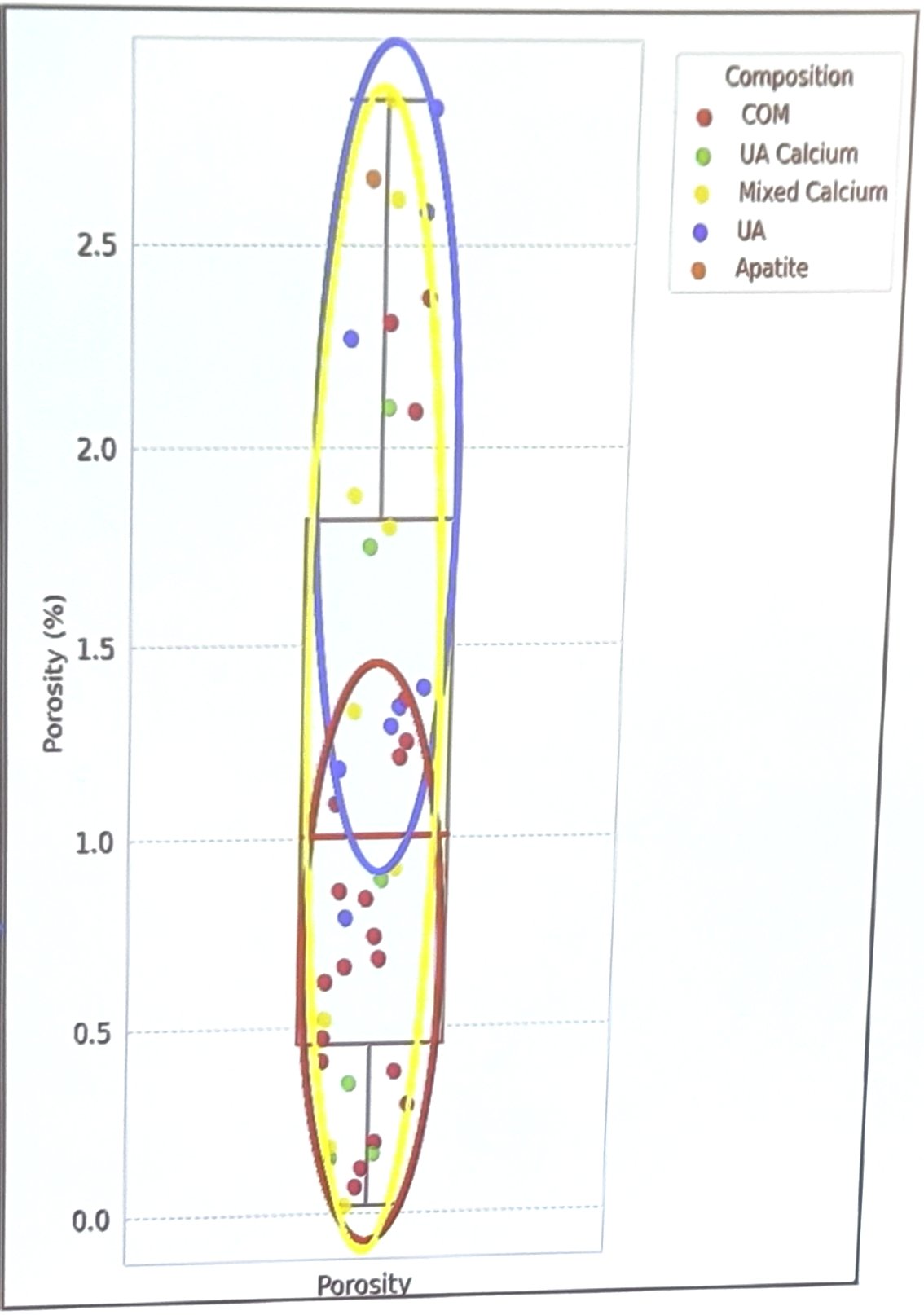
Figure 3. Porosity of different kidney stones
Gavin concluded that this study demonstrated that porosity is associated with lower density, HUs, and Vickers hardness. With that being said, HUs may be used as a predictor of how susceptible a kidney stone is to stone fragmentation during laser lithotripsy.
At the conclusion of the podium presentation, an audience member asked Gavin if his team had stone fragments containing both uric acid and calcium oxalate. Gavin provided a clear response that his team had a subset of stones with this characteristic, of which these stones had a uric acid core with a COM periphery. He further stated that this spatial information is relevant for how a surgeon would proceed for stone fragmentation and is even more important when comparing to how the use of Fourier Transform Infrared (FTIR) spectroscopy for kidney stone composition analysis leads to a loss in the spatial composition of the stone from overall stone destruction.
Presented by: Gavin Hughes, MS-2, University of Toronto, Toronto, Canada during the 2026 American Urological Association (AUA) Annual Meeting, May 15 – May 18, 2026, Washington, D.C.
Written by: Victor Pham, B.S., University of California Irvine, @victorpham01 on X during the American Urological Association (AUA) 2026 Annual Meeting, Washington, DC, Fri, May 15 – Mon, May 18, 2026.