(UroToday.com) During the Saturday morning plenary session, Dr. Joseph C. Liao delivered a state-of-the-art lecture on Computer Vision in Endourology, highlighting how artificial intelligence (AI) and computer vision technologies are beginning to reshape multiple aspects of modern urologic surgery. Throughout the presentation, Dr. Liao discussed the growing applications of AI-assisted image analysis across cystoscopy, ureteroscopy, robotic surgery, surgical education, and intraoperative decision support, emphasizing that the field is gradually moving from proof-of-concept research toward real-time clinical integration.
Dr. Liao began by explaining why endourology represents a particularly natural fit for computer vision applications. Because nearly all endourologic procedures are performed through digital endoscopic visualization displayed on monitors, the specialty already generates large amounts of visual data that can be analyzed computationally. He described computer vision as a branch of AI focused on automated interpretation of images and videos, including tasks such as object detection, image classification, segmentation, and spatial reconstruction. According to Dr. Liao, recent advances in convolutional neural networks (CNNs), improved graphics processing capabilities, and the availability of large annotated datasets have significantly accelerated progress in this area over recent years.
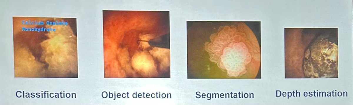
Several foundational computer vision tasks were reviewed during the lecture. Classification allows AI systems to identify what structure is being visualized, while object detection determines where abnormalities are located within the image. Segmentation further refines this process by outlining structures at the pixel level, enabling precise delineation of tissue or stone boundaries. Dr. Liao also discussed more advanced spatial reconstruction techniques capable of generating depth estimation and three-dimensional understanding during endoscopic procedures, potentially improving surgical visualization and navigation.
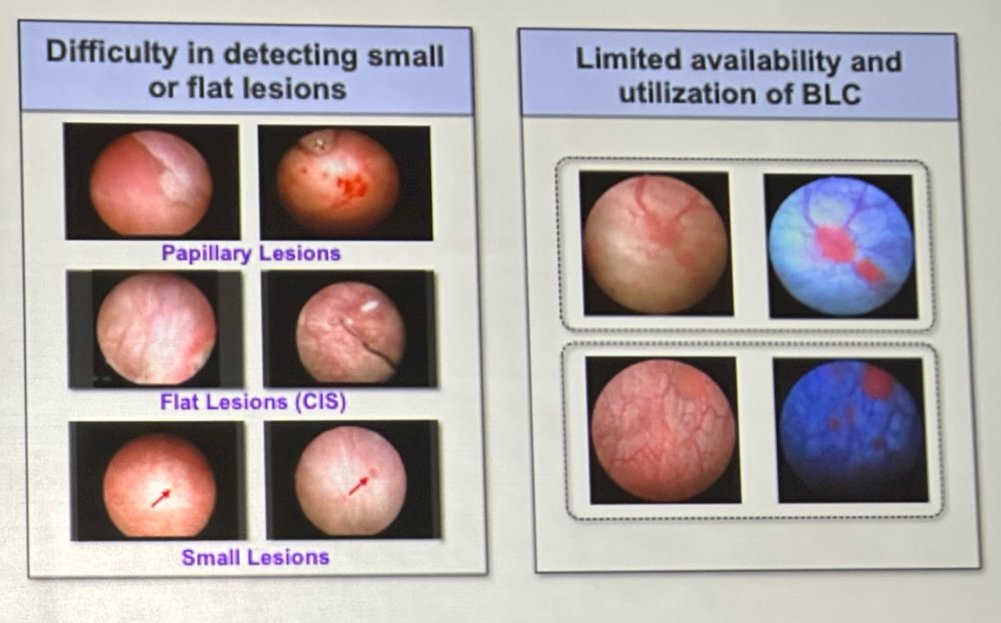
One of the major clinical applications discussed was bladder tumor detection during cystoscopy. Dr. Liao noted that conventional white-light cystoscopy remains highly operator dependent and may occasionally miss small papillary tumors or flat lesions such as carcinoma in situ. To address these limitations, several groups have developed CNN-based models trained using pathologically confirmed cystoscopic images and videos. Early studies demonstrated encouraging performance, with some systems achieving sensitivities and specificities approaching expert-level interpretation.
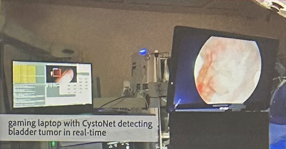
Importantly, the field is now beginning to move beyond retrospective image analysis toward real-time intraoperative implementation. Dr. Liao presented work from his group involving a computer vision platform integrated directly into a cystoscopy tower using a portable gaming laptop capable of performing live tumor detection with clinically acceptable latency. He also reviewed similar efforts from international groups developing real-time AI-assisted cystoscopy systems, further supporting the feasibility of clinical deployment during active procedures.

Applications in upper tract urothelial carcinoma were also highlighted. Researchers have developed automated ureteroscopic tumor segmentation systems trained using thousands of manually annotated video frames. Some models demonstrated excellent discrimination performance while maintaining near real-time processing speeds, suggesting that AI-assisted upper tract tumor detection during ureteroscopy may become increasingly feasible in the future.
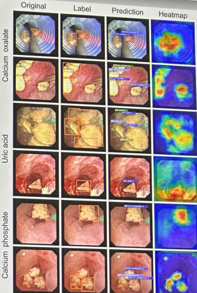
Another major focus of the lecture involved computer vision applications for kidney stone analysis. Dr. Liao explained that laser lithotripsy frequently fragments stones extensively, which may limit the ability to obtain adequate specimens for formal chemical analysis. However, intraoperative knowledge of stone composition may help optimize laser settings and guide postoperative metabolic management. Early work from the University of Michigan demonstrated that AI models trained on digital stone photographs could classify major stone types with promising accuracy. Subsequent studies expanded this concept using ureteroscopic video frames rather than still images while also incorporating automated stone segmentation.
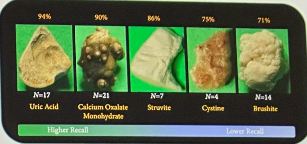
Dr. Liao also reviewed more advanced multi-task AI systems capable of simultaneously performing stone detection, composition classification, stone sizing, and residual fragment quantification in real time. Some models demonstrated strong performance across multiple stone compositions, including calcium oxalate and calcium phosphate stones. Particularly notable was the ability of these systems to quantify residual fragments smaller than 1–2 mm and correlate those findings with future reoperation risk, representing one of the first examples of computer vision data being directly linked to clinically meaningful postoperative outcomes in endourology.
The lecture additionally explored the growing role of computer vision in surgical education and skill assessment. Historically, evaluation of operative performance has relied heavily on subjective assessment. Using automated video analysis, investigators were able to objectively assess ureteroscopic performance metrics such as scope stability, stone localization, and laser targeting efficiency. These systems successfully differentiated expert surgeons from trainees and may ultimately provide valuable tools for procedural training and competency assessment.
In robotic surgery, Dr. Liao discussed how computer vision applications have progressed even further. He reviewed pioneering work utilizing AI-based analysis of robotic-assisted radical prostatectomy videos by dividing procedures into discrete surgical gestures during nerve-sparing dissection. By analyzing tens of thousands of annotated surgical gestures, investigators identified specific operative maneuvers associated with improved postoperative erectile function recovery. These findings demonstrated how AI-driven surgical video analysis may eventually provide an objective assessment of surgical quality while also helping predict patient functional outcomes.
In his concluding remarks, Dr. Liao emphasized that the field remains in an important transitional phase as researchers work to move computer vision systems from experimental studies into real-world clinical practice. He noted that several key challenges still need to be addressed, including human factor testing, multicenter dataset standardization, validation across diverse surgical environments, regulatory oversight, and cost considerations. Nevertheless, he expressed strong optimism that computer vision technologies will become an increasingly important component of future urologic surgery.
In summary, the presentation provided an insightful overview of how AI-powered computer vision is beginning to influence multiple areas of endourology, from tumor detection and kidney stone analysis to surgical education and robotic surgery. As these technologies continue to mature, they may significantly change how urologists visualize disease, perform procedures, assess technical performance, and ultimately improve patient outcomes.
Presented by: Joseph C. Liao, MD, Department of Urology, Stanford University School of Medicine, Stanford, CA
Written by: Kantapon Tangwiwat, MD, Research Fellow, Department of Urology, University of California, Irvine, CA during the 2026 American Urological Association (AUA) Annual Meeting, May 15– 18, 2026, Washington D.C.