(UroToday.com) The 2025 ASTRO annual meeting featured a localized prostate cancer session and a presentation by Lily Nguyen discussing PURE-MRI, an international study assessing physician accuracy in delineating the prostate and urethra on prostate MRI. There is increasing use of MRI for prostate cancer treatment planning, including in the FLAME trial1 using focal boost to visible MRI lesions showing improved outcomes.
Precise delineation of genitourinary structures during prostate cancer care is critical to optimize treatment delivery while minimizing toxicity and injury; however, it is unclear how well physicians identify these structures. The Prostate and UREthra on MRI (PURE-MRI) study is an international, prospective study to assess physicians’ accuracy in segmenting the prostate and urethra on MRI, and to evaluate auto-segmentation artificial intelligence tools.
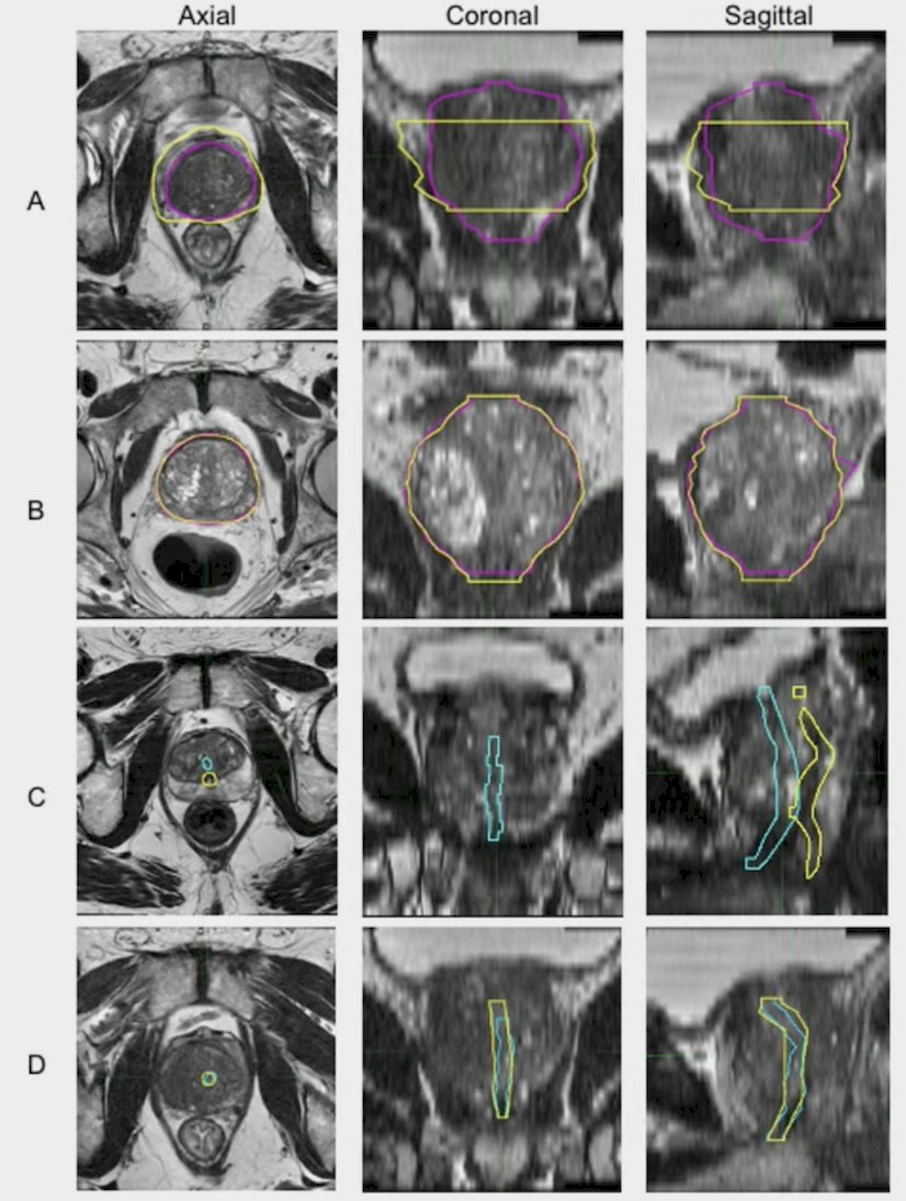
Physicians who diagnose or treat prostate cancer were invited to contour the prostate and urethra on up to 4 patient cases using standard T2-weighted MRI (all planes). These contours were compared to reference consensus segmentations produced by a multidisciplinary panel of experts. The performance of a validated prostate auto-segmentation AI tool was also evaluated. The investigators assessed contour accuracy with Dice similarity coefficient, deviation from boundary (mm), absolute volume difference (%), and overlap (%):
A mixed effects model was used to evaluate potential associations between contour performance and physician specialty, genitourinary focus, or clinical experience.
59 radiation oncologists, radiologists, and urologists (from 11 countries) created a total of 212m structure segmentations (108 prostate, 104 urethra). Dice similarity coefficient for the prostate, reported as median, was 0.92 (min 0.90, max 0.94) for physicians, with no clear effect of clinical experience or focus. Maximum deviation inside (cutting into expert contour), maximum deviation outside (extending beyond expert contour), and mean deviation (per case) from the reference prostate were 3.4 mm (min 1.0, max 12.4), 5.3 mm (min 2.4, max 7.0), and 1.6 mm (min 1.3, max 17.3), respectively. By comparison, the prostate auto-segmentation tool had Dice similarity coefficient, max deviation inside, max distance outside, and mean deviation per case of 0.95 (min 0.94, max 0.96), 3 mm, 3.9 mm (min 3.1, max 4.9), and 1.2 mm (min 1.1, max 1.5), respectively:

Physician performance was considerably worse for the urethra, with Dice similarity coefficient of 0.33 (min 0.03, max 0.69). Artificial intelligence tools contoured the urethra more accurately than physicians:
The linear mixed effects model for prostate Dice score showed that there was no major effect of physician specialty, and that attending physicians were not more accurate than trainees:
Lily Nguyen concluded her presentation discussing PURE-MRI, an international study assessing physician accuracy in delineating the prostate and urethra on prostate MRI, with the following take-home points:
- Physicians contour the prostate on MRI with reasonably high accuracy (despite some significant errors), but their urethra contours are both highly variable and inaccurate
- Contouring is not a major barrier to adopting MRI for prostate cancer radiotherapy (trainees do as well as experienced specialists)
- Artificial intelligence tools contour the prostate at least as well as physicians and contour the urethra more accurately than physicians
Presented by: Lily Nguyen, UCSD Medical School, La Jolla, CA
Written by: Zachary Klaassen, MD, MSc – Urologic Oncologist, Associate Professor of Urology, Georgia Cancer Center, Wellstar MCG Health, @zklaassen_md on Twitter during the 2025 American Society for Radiation Oncology (ASTRO) Annual Meeting, San Francisco, CA, Sat, Sept 27 – Wed, Oct 1, 2025.
Reference: