(UroToday.com) The 2025 American Society of Clinical Oncology (ASCO) Annual Meeting held in Chicago, IL between May 30 and June 3 was host to the Poster Session: Genitourinary Cancer - Prostate, Testicular, and Penile Cancer. Luke Nordquist, MD, FACP, presented Poster 5102: COBRA: Assessment of the efficacy of 64Cu-SAR-bisPSMA using histopathology and standard of care imaging as reference standard in patients with biochemical recurrence of prostate cancer following definitive therapy.
Approximately 20%–40% of patients with prostate cancer (PCa) experience biochemical recurrence (BCR) within 10 years of initial treatment, typically signaled by a rising PSA level. Early detection and accurate staging are essential for guiding subsequent management. Prostate-specific membrane antigen (PSMA) has emerged as a critical molecular target for imaging in the setting of recurrent PCa. Although currently approved PSMA PET agents provide high specificity, their sensitivity remains limited—highlighting an ongoing need for next-generation imaging tools to improve staging accuracy in this context.1
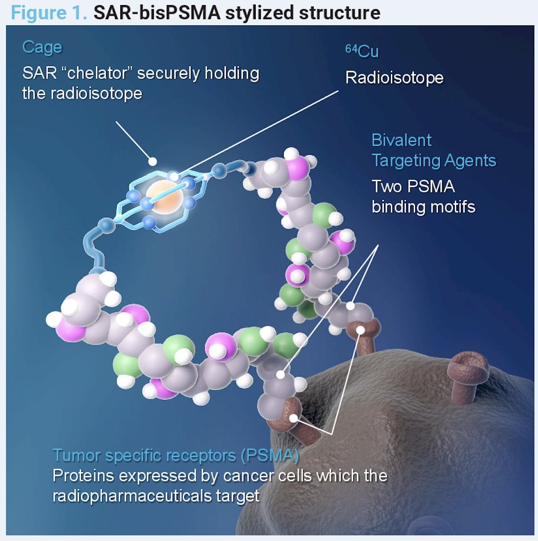
Accurate staging of PCa is critical to informing optimal treatment strategies. ⁶⁴Cu-SAR-bisPSMA is a novel bivalent PSMA-targeting radiotracer designed to enhance imaging sensitivity and tumor localization. As illustrated in the figure below, this agent features a unique dual PSMA-binding motif enhancing tumor affinity and a SAR “chelator” cage that securely holds the ⁶⁴Cu radioisotope.
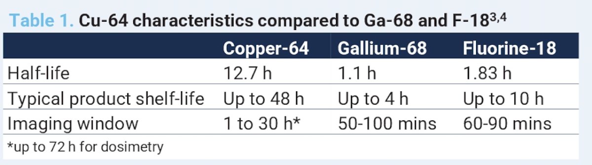
Moreover, the longer half-life of copper-64 (12.7 hours vs. <2 hours for ¹⁸F and ⁶⁸Ga) contribute to improved pharmacokinetics, enabling higher tumor uptake and enhanced lesion detection.
Clinical evidence has shown that ⁶⁴Cu-SAR-bisPSMA achieves 2- to 3-fold higher tumor uptake on same-day imaging and enables detection of additional prostate cancer lesions compared to currently approved PSMA PET agents.2,3
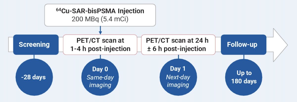
COBRA was a Phase 1/2 study (NCT05249127) evaluating the safety and efficacy of ⁶⁴Cu-SAR-bisPSMA (200 MBq) in patients with PCa experiencing biochemical recurrence following definitive therapy. Eligible patients had histologically confirmed adenocarcinoma of the prostate, rising or detectable PSA levels suggestive of recurrence, and negative or equivocal findings for PCa on conventional imaging within 60 days prior to study enrollment. The trial specifically targeted a population with limited localization options using standard-of-care imaging, aiming to assess the added diagnostic value of this novel PSMA-targeted radiotracer.
PET/CT imaging was performed on Day 0 (1–4 hours post-dose) and Day 1 (24±6 hours post-dose), with images reviewed by three blinded central readers. PET/CT findings were compared against a composite reference standard, which included histopathology, SOC imaging interpreted by two independent readers, and PSA response following focal therapy.
Primary objective and endpoints are summarized in the table below:
PET imaging with ⁶⁴Cu-SAR-bisPSMA was evaluated using a hierarchical reference standard framework. All PET/CT scans were interpreted independently by three blinded, central readers. The imaging findings were compared against a composite reference standard, adjudicated by an independent, blinded central expert panel.
This composite standard incorporated up to three levels of evidence:- Evaluable histopathology from biopsy or surgery, when available;
- Conventional imaging, if histopathology was unavailable, inconclusive, or negative;
- Follow-up PSMA PET or confirmed PSA response following salvage focal therapy (without concomitant ADT), defined as a ≥50% decline in PSA from baseline confirmed within four weeks, per PCWG3 criteria.
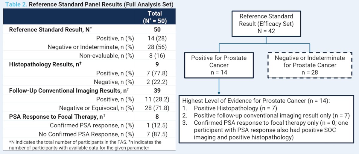
A total of 52 participants were enrolled and received ⁶⁴Cu-SAR-bisPSMA PET imaging, comprising the safety analysis set. Of these, 50 patients continued with follow-up and constituted the full analysis set. Eight patients lacked sufficient reference standard data, resulting in 42 patients being included in the efficacy analysis set. A summary of the reference standard efficacy set and the full analysis set is illustrated below.
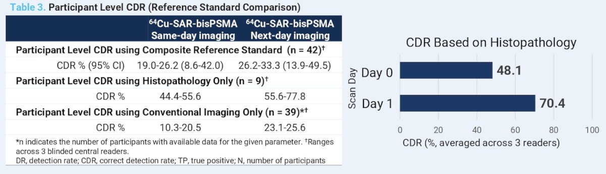
The correct detection rate (CDR) of ⁶⁴Cu-SAR-bisPSMA was higher on next-day imaging and when histopathology alone was used as the reference standard, compared to same-day imaging and validation using conventional follow-up imaging. Notably, CDR was significantly higher when benchmarked against histopathology (the gold standard), CDR: 44.4–55.6% on Day 0 and 55.6–77.8% on Day 1. Notably, in cases where biopsies were performed, histopathology confirmed prostate cancer in up to 78% of lesions identified by ⁶⁴Cu-SAR-bisPSMA PET.
When CDR was evaluated against the composite reference standard (which included histopathology, conventional imaging, and PSA response), CDR ranged from 19.0% to 26.2% on Day 0 and improved to 26.2% to 33.3% on Day 1. CDR was impacted by the high number of lesions that were detected but could not be biopsied for clinical reasons, as well as by the limited sensitivity of conventional imaging methods used for validation.
Detection rates with ⁶⁴Cu-SAR-bisPSMA were higher on next-day imaging compared to same-day imaging. On average across the three blinded readers, 71% of participants had a positive scan on next-day imaging versus 53% on same-day imaging representing a 34% relative increase in detection rate.
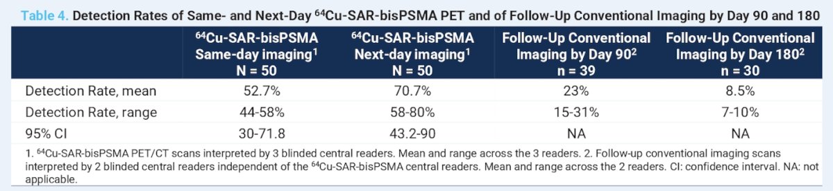
Quantitative analysis comparing same-day and next-day ⁶⁴Cu-SAR-bisPSMA imaging demonstrated substantial increases in tracer uptake and lesion contrast. SUVmean, SUVmax, and tumor-to-background ratio (TBR) were assessed in up to 25 lesions per patient across all scans. Averaged across three blinded readers, next-day imaging showed markedly higher values: SUVmean increased from 6.6–9.9 (same-day) to 14.7–15.8, and SUVmax rose from 13.9–14.0 to 22.2–33.4. TBR improved significantly, from 23.2–25.4 on same-day imaging to 118.1–181.7 on next-day imaging representing more than a fivefold increase. Notably, lesion SUVmax and SUVmean were >80% higher on next-day imaging, highlighting the added diagnostic value of delayed acquisition.
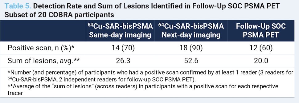
Follow-up PSMA PET imaging was obtained in 20 participants, 13 with ⁶⁸Ga-PSMA-11 and 7 with ¹⁸F-DCFPyL. The median interval between same-day ⁶⁴Cu-SAR-bisPSMA imaging and follow-up SOC PSMA PET was 73.5 days. In this subset of patients, ⁶⁴Cu-SAR-bisPSMA demonstrated a higher detection rate than PSMA agents, even when the comparator scan was performed up to 180 days later. These findings suggest that ⁶⁴Cu-SAR-bisPSMA can identify lesions as early as 29 days and in some cases more than six months prior to detection by conventional PSMA PET agents.
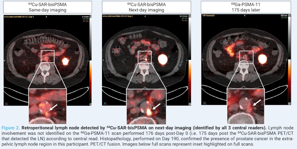
Dr. Nordquist highlighted a representative case in which ⁶⁴Cu-SAR-bisPSMA PET/CT identified pelvic nodal involvement that had not been detected on a subsequent ⁶⁸Ga-PSMA-11 PET/CT performed 175 days later. On next-day imaging, a retroperitoneal lymph node was identified by all three central readers using ⁶⁴Cu-SAR-bisPSMA. This lesion was not visualized on the ⁶⁸Ga-PSMA-11 scan obtained 176 days after baseline (Day 0). Histopathologic confirmation on Day 190 verified the presence of prostate cancer in the extra-pelvic lymph node.
Dr. Nordquist concluded with the following key messages from the COBRA study:
- ⁶⁴Cu-SAR-bisPSMA demonstrated high detection capability in patients with biochemical recurrence, identifying lesions in up to 80% of participants who had negative or equivocal findings on baseline conventional imaging.
- Next-day imaging outperformed same-day imaging, with a higher proportion of participants showing positive findings on delayed scans.
- Correct detection rates were significantly higher when histopathology was used as the verification standard compared to conventional imaging, underscoring the limitations of less-sensitive reference methods.
- In a subset of patients with follow-up SOC PSMA PET scans up to 180 days later, more lesions were detected using ⁶⁴Cu-SAR-bisPSMA, suggesting it may enable earlier lesion identification than currently approved PSMA agents.
- These findings have important clinical implications, as earlier and more accurate lesion detection in patients with BCR can directly influence treatment decisions and management strategies.
- The upcoming registrational Phase III AMPLIFY trial will further evaluate ⁶⁴Cu-SAR-bisPSMA PET in patients with BCR following definitive therapy.
Presented by: Luke Nordquist, MD, FACP, CEO of Urology Cancer Center & GU Research, CEO XCancer Research Network, Chairman XCancer Foundation. Omaha, Nebraska.
Written by: Julian Chavarriaga, MD – Urologic Oncologist at Cancer Treatment and Research Center (CTIC) via Society of Urologic Oncology (SUO) Fellow at The University of Toronto. @chavarriagaj on Twitter during the American Society of Clinical Oncology (ASCO) 2025 Annual Meeting, Chicago, IL, Fri, May 30 – Tues, Jun 3, 2025.
References:- Pak S, You D, Jeong IG, Kim YS, Hong JH, Kim CS, Ahn H. Time to biochemical relapse after radical prostatectomy and efficacy of salvage radiotherapy in patients with prostate cancer. Int J Clin Oncol. 2019 Oct;24(10):1238-1246. doi: 10.1007/s10147-019-01463-5. Epub 2019 May 13. PMID: 31087170.
- Lengyelova H, Violet J, Hofman MS, et al. First-in-human evaluation of ⁶⁴Cu-SAR-bisPSMA in patients with metastatic prostate cancer. Presented at: ASCO Annual Meeting; 2023.
- Nordquist L, Emmett L, Shah A, et al. ⁶⁴Cu-SAR-bisPSMA PET detects more lesions than ⁶⁸Ga-PSMA-11: Results from a prospective intra-individual comparison study. Presented at: ASCO Annual Meeting; 2024.